Abeer A Alhazzani, Mohanned F Tobaigy, Munirah I Aldofyan, Abdulrahman F AlBloushi
{"title":"周围视网膜下肿块合并坏死性前巩膜炎一例肉芽肿合并多血管炎。","authors":"Abeer A Alhazzani, Mohanned F Tobaigy, Munirah I Aldofyan, Abdulrahman F AlBloushi","doi":"10.4103/meajo.meajo_180_22","DOIUrl":null,"url":null,"abstract":"<p><p>Anterior scleritis is rarely diagnosed with a peripheral amelanotic subretinal mass. We reported a rare case of a 31-year-old woman who was referred for suspected left eye choroidal melanoma. The patient had granulomatosis with polyangiitis with a history of treated left eye necrotizing anterior scleritis. Her left eye examination revealed 20/60 vision, superotemporal diffuse scleral injection, and thinning. Dilated fundus examination of the left eye showed a large peripheral amelanotic subretinal mass below the area of anterior scleritis, optic disc hyperemia, and subretinal fluid. The patient was successfully treated with intravenous methylprednisolone, rituximab infusions, and oral methotrexate. Two months after treatment, her vision improved to 20/20, with inactive anterior scleritis and a significant reduction in the subretinal mass with complete resolution of optic disc hyperemia and subretinal fluid. High index of suspicion of this atypical presentation of anterior scleritis is important to avoid aggressive modalities of treatment.</p>","PeriodicalId":18740,"journal":{"name":"Middle East African Journal of Ophthalmology","volume":"29 3","pages":"159-162"},"PeriodicalIF":0.3000,"publicationDate":"2022-07-01","publicationTypes":"Journal Article","fieldsOfStudy":null,"isOpenAccess":false,"openAccessPdf":"https://www.ncbi.nlm.nih.gov/pmc/articles/PMC10319073/pdf/","citationCount":"1","resultStr":"{\"title\":\"Peripheral Subretinal Mass Complicating Necrotizing Anterior Scleritis in a Patient with Granulomatosis with Polyangiitis.\",\"authors\":\"Abeer A Alhazzani, Mohanned F Tobaigy, Munirah I Aldofyan, Abdulrahman F AlBloushi\",\"doi\":\"10.4103/meajo.meajo_180_22\",\"DOIUrl\":null,\"url\":null,\"abstract\":\"<p><p>Anterior scleritis is rarely diagnosed with a peripheral amelanotic subretinal mass. We reported a rare case of a 31-year-old woman who was referred for suspected left eye choroidal melanoma. The patient had granulomatosis with polyangiitis with a history of treated left eye necrotizing anterior scleritis. Her left eye examination revealed 20/60 vision, superotemporal diffuse scleral injection, and thinning. Dilated fundus examination of the left eye showed a large peripheral amelanotic subretinal mass below the area of anterior scleritis, optic disc hyperemia, and subretinal fluid. The patient was successfully treated with intravenous methylprednisolone, rituximab infusions, and oral methotrexate. Two months after treatment, her vision improved to 20/20, with inactive anterior scleritis and a significant reduction in the subretinal mass with complete resolution of optic disc hyperemia and subretinal fluid. High index of suspicion of this atypical presentation of anterior scleritis is important to avoid aggressive modalities of treatment.</p>\",\"PeriodicalId\":18740,\"journal\":{\"name\":\"Middle East African Journal of Ophthalmology\",\"volume\":\"29 3\",\"pages\":\"159-162\"},\"PeriodicalIF\":0.3000,\"publicationDate\":\"2022-07-01\",\"publicationTypes\":\"Journal Article\",\"fieldsOfStudy\":null,\"isOpenAccess\":false,\"openAccessPdf\":\"https://www.ncbi.nlm.nih.gov/pmc/articles/PMC10319073/pdf/\",\"citationCount\":\"1\",\"resultStr\":null,\"platform\":\"Semanticscholar\",\"paperid\":null,\"PeriodicalName\":\"Middle East African Journal of Ophthalmology\",\"FirstCategoryId\":\"1085\",\"ListUrlMain\":\"https://doi.org/10.4103/meajo.meajo_180_22\",\"RegionNum\":0,\"RegionCategory\":null,\"ArticlePicture\":[],\"TitleCN\":null,\"AbstractTextCN\":null,\"PMCID\":null,\"EPubDate\":\"\",\"PubModel\":\"\",\"JCR\":\"Q4\",\"JCRName\":\"OPHTHALMOLOGY\",\"Score\":null,\"Total\":0}","platform":"Semanticscholar","paperid":null,"PeriodicalName":"Middle East African Journal of Ophthalmology","FirstCategoryId":"1085","ListUrlMain":"https://doi.org/10.4103/meajo.meajo_180_22","RegionNum":0,"RegionCategory":null,"ArticlePicture":[],"TitleCN":null,"AbstractTextCN":null,"PMCID":null,"EPubDate":"","PubModel":"","JCR":"Q4","JCRName":"OPHTHALMOLOGY","Score":null,"Total":0}
Peripheral Subretinal Mass Complicating Necrotizing Anterior Scleritis in a Patient with Granulomatosis with Polyangiitis.
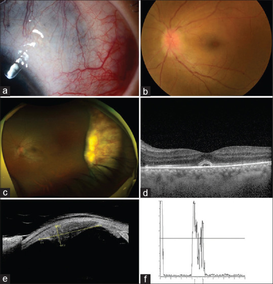
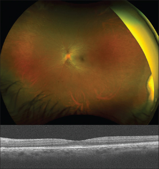
Anterior scleritis is rarely diagnosed with a peripheral amelanotic subretinal mass. We reported a rare case of a 31-year-old woman who was referred for suspected left eye choroidal melanoma. The patient had granulomatosis with polyangiitis with a history of treated left eye necrotizing anterior scleritis. Her left eye examination revealed 20/60 vision, superotemporal diffuse scleral injection, and thinning. Dilated fundus examination of the left eye showed a large peripheral amelanotic subretinal mass below the area of anterior scleritis, optic disc hyperemia, and subretinal fluid. The patient was successfully treated with intravenous methylprednisolone, rituximab infusions, and oral methotrexate. Two months after treatment, her vision improved to 20/20, with inactive anterior scleritis and a significant reduction in the subretinal mass with complete resolution of optic disc hyperemia and subretinal fluid. High index of suspicion of this atypical presentation of anterior scleritis is important to avoid aggressive modalities of treatment.
期刊介绍:
The Middle East African Journal of Ophthalmology (MEAJO), published four times per year in print and online, is an official journal of the Middle East African Council of Ophthalmology (MEACO). It is an international, peer-reviewed journal whose mission includes publication of original research of interest to ophthalmologists in the Middle East and Africa, and to provide readers with high quality educational review articles from world-renown experts. MEAJO, previously known as Middle East Journal of Ophthalmology (MEJO) was founded by Dr Akef El Maghraby in 1993.


 分享
分享
 求助内容:
求助内容: 应助结果提醒方式:
 扫码关注我们
