Fardad Jabbary Aslany, Kimberly McBain, Liang Chen, Jeremy O'Brien, Geoffroy P J C Noel
{"title":"解剖过程中与增强现实耳机配合使用的尸检前和尸检后尸体图像的比较。","authors":"Fardad Jabbary Aslany, Kimberly McBain, Liang Chen, Jeremy O'Brien, Geoffroy P J C Noel","doi":"10.1007/s00276-023-03239-z","DOIUrl":null,"url":null,"abstract":"<p><strong>Purpose: </strong>Medical training has undergone many transformations to incorporate diagnostic imaging along side anatomical education. Post-mortem computed tomography (CT) scanning of body donors prior to dissection has been proposed. However, it poses challenges secondary to the embalming process and other post-mortem physiological changes that significantly alter the imaging quality. The purposes of this study were to compare the accuracy of pathology identification on pre- and post-mortem CT scans of body donors and to assess the integration of those scans in a dissection-based course, where these images were overlaid onto body donors using augmented reality (AR).</p><p><strong>Methods: </strong>Participants in this study included 35 fourth year medical students, 5 radiology residents and 3 radiologists. A convergent, parallel mixed methods design was employed with quantitative measures that included statistical analyses of a double-blinded comparison of pathological lesions recognition, on both image sets, the group responses to a study participant survey and the login access data from imaging repository. The study also included qualitative analysis of post-elective structured interviews.</p><p><strong>Results: </strong>The double-blinded comparison revealed that staff radiologists can only identify, on post-mortem images, 54.8% of the pathologies that they were able to detect on the pre-mortem scans. Analyses of the surveys and login access data reveal that 60% of radiology residents and 56% of students preferred pre-mortem scans and used those scans more often than post-mortem scans (67 access vs 36, respectively). However, post-mortem scans were significantly preferred when used to overlay onto body donors using AR (p = 0.0047).</p><p><strong>Conclusion: </strong>These results show that post-mortem imaging can be valuable alongside pre-mortem imaging, as they represent the most concordance between the anatomical structures and pathologies seen on the images and what is being dissected.</p>","PeriodicalId":49296,"journal":{"name":"Surgical and Radiologic Anatomy","volume":" ","pages":"1311-1319"},"PeriodicalIF":1.2000,"publicationDate":"2023-10-01","publicationTypes":"Journal Article","fieldsOfStudy":null,"isOpenAccess":false,"openAccessPdf":"","citationCount":"0","resultStr":"{\"title\":\"Comparison between pre-mortem and post-mortem cadaveric images for use with augmented reality headsets during dissection.\",\"authors\":\"Fardad Jabbary Aslany, Kimberly McBain, Liang Chen, Jeremy O'Brien, Geoffroy P J C Noel\",\"doi\":\"10.1007/s00276-023-03239-z\",\"DOIUrl\":null,\"url\":null,\"abstract\":\"<p><strong>Purpose: </strong>Medical training has undergone many transformations to incorporate diagnostic imaging along side anatomical education. Post-mortem computed tomography (CT) scanning of body donors prior to dissection has been proposed. However, it poses challenges secondary to the embalming process and other post-mortem physiological changes that significantly alter the imaging quality. The purposes of this study were to compare the accuracy of pathology identification on pre- and post-mortem CT scans of body donors and to assess the integration of those scans in a dissection-based course, where these images were overlaid onto body donors using augmented reality (AR).</p><p><strong>Methods: </strong>Participants in this study included 35 fourth year medical students, 5 radiology residents and 3 radiologists. A convergent, parallel mixed methods design was employed with quantitative measures that included statistical analyses of a double-blinded comparison of pathological lesions recognition, on both image sets, the group responses to a study participant survey and the login access data from imaging repository. The study also included qualitative analysis of post-elective structured interviews.</p><p><strong>Results: </strong>The double-blinded comparison revealed that staff radiologists can only identify, on post-mortem images, 54.8% of the pathologies that they were able to detect on the pre-mortem scans. Analyses of the surveys and login access data reveal that 60% of radiology residents and 56% of students preferred pre-mortem scans and used those scans more often than post-mortem scans (67 access vs 36, respectively). However, post-mortem scans were significantly preferred when used to overlay onto body donors using AR (p = 0.0047).</p><p><strong>Conclusion: </strong>These results show that post-mortem imaging can be valuable alongside pre-mortem imaging, as they represent the most concordance between the anatomical structures and pathologies seen on the images and what is being dissected.</p>\",\"PeriodicalId\":49296,\"journal\":{\"name\":\"Surgical and Radiologic Anatomy\",\"volume\":\" \",\"pages\":\"1311-1319\"},\"PeriodicalIF\":1.2000,\"publicationDate\":\"2023-10-01\",\"publicationTypes\":\"Journal Article\",\"fieldsOfStudy\":null,\"isOpenAccess\":false,\"openAccessPdf\":\"\",\"citationCount\":\"0\",\"resultStr\":null,\"platform\":\"Semanticscholar\",\"paperid\":null,\"PeriodicalName\":\"Surgical and Radiologic Anatomy\",\"FirstCategoryId\":\"3\",\"ListUrlMain\":\"https://doi.org/10.1007/s00276-023-03239-z\",\"RegionNum\":4,\"RegionCategory\":\"医学\",\"ArticlePicture\":[],\"TitleCN\":null,\"AbstractTextCN\":null,\"PMCID\":null,\"EPubDate\":\"2023/9/12 0:00:00\",\"PubModel\":\"Epub\",\"JCR\":\"Q3\",\"JCRName\":\"ANATOMY & MORPHOLOGY\",\"Score\":null,\"Total\":0}","platform":"Semanticscholar","paperid":null,"PeriodicalName":"Surgical and Radiologic Anatomy","FirstCategoryId":"3","ListUrlMain":"https://doi.org/10.1007/s00276-023-03239-z","RegionNum":4,"RegionCategory":"医学","ArticlePicture":[],"TitleCN":null,"AbstractTextCN":null,"PMCID":null,"EPubDate":"2023/9/12 0:00:00","PubModel":"Epub","JCR":"Q3","JCRName":"ANATOMY & MORPHOLOGY","Score":null,"Total":0}
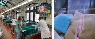
Comparison between pre-mortem and post-mortem cadaveric images for use with augmented reality headsets during dissection.
Purpose: Medical training has undergone many transformations to incorporate diagnostic imaging along side anatomical education. Post-mortem computed tomography (CT) scanning of body donors prior to dissection has been proposed. However, it poses challenges secondary to the embalming process and other post-mortem physiological changes that significantly alter the imaging quality. The purposes of this study were to compare the accuracy of pathology identification on pre- and post-mortem CT scans of body donors and to assess the integration of those scans in a dissection-based course, where these images were overlaid onto body donors using augmented reality (AR).
Methods: Participants in this study included 35 fourth year medical students, 5 radiology residents and 3 radiologists. A convergent, parallel mixed methods design was employed with quantitative measures that included statistical analyses of a double-blinded comparison of pathological lesions recognition, on both image sets, the group responses to a study participant survey and the login access data from imaging repository. The study also included qualitative analysis of post-elective structured interviews.
Results: The double-blinded comparison revealed that staff radiologists can only identify, on post-mortem images, 54.8% of the pathologies that they were able to detect on the pre-mortem scans. Analyses of the surveys and login access data reveal that 60% of radiology residents and 56% of students preferred pre-mortem scans and used those scans more often than post-mortem scans (67 access vs 36, respectively). However, post-mortem scans were significantly preferred when used to overlay onto body donors using AR (p = 0.0047).
Conclusion: These results show that post-mortem imaging can be valuable alongside pre-mortem imaging, as they represent the most concordance between the anatomical structures and pathologies seen on the images and what is being dissected.
期刊介绍:
Anatomy is a morphological science which cannot fail to interest the clinician. The practical application of anatomical research to clinical problems necessitates special adaptation and selectivity in choosing from numerous international works. Although there is a tendency to believe that meaningful advances in anatomy are unlikely, constant revision is necessary. Surgical and Radiologic Anatomy, the first international journal of Clinical anatomy has been created in this spirit.
Its goal is to serve clinicians, regardless of speciality-physicians, surgeons, radiologists or other specialists-as an indispensable aid with which they can improve their knowledge of anatomy. Each issue includes: Original papers, review articles, articles on the anatomical bases of medical, surgical and radiological techniques, articles of normal radiologic anatomy, brief reviews of anatomical publications of clinical interest.
Particular attention is given to high quality illustrations, which are indispensable for a better understanding of anatomical problems.
Surgical and Radiologic Anatomy is a journal written by anatomists for clinicians with a special interest in anatomy.


 分享
分享
 求助内容:
求助内容: 应助结果提醒方式:
 扫码关注我们
