{"title":"病理性骨化引起尺骨神经病变1例。","authors":"Kunc Vojtěch, Kachlík David, Humhej Ivan","doi":"10.1007/s00276-023-03217-5","DOIUrl":null,"url":null,"abstract":"<p><strong>Purpose: </strong>Cubital tunnel syndrome is a well-described entity with many reported etiologies and anatomical compression sites. Accessory ossicles of either traumatic or congenital origin might occur around the elbow joint. Only one case reporting such ossicles compressing the ulnar nerve exists in previous literature. We aim to present this entity with a detailed description of the patient history and treatment.</p><p><strong>Case report: </strong>We report a case of 30-year-old female presenting with classical signs of cubital tunnel syndrome-positive Wartenberg's and Froment's signs, hypoesthesia in the fourth and fifth finger with decreased finger duction strength but without gross hypotrophy of interosseous and hypothenar muscles. Tinel's sign was positive over the ulnar sulcus and an accessory ossicle was found on the elbow radiograph within the ulnar sulcus. The first signs of calcification in this patient were reported 6 years prior in a follow-up after the dislocation of her elbow joint following a bike accident. The EMG confirmed ulnar nerve neuropathy in the elbow area. The ossicle was extirpated, the ulnar nerve was decompressed in the ulnar sulcus in a standard manner and the symptoms quickly resolved. The patient has been regularly visiting our outpatient clinic for the next 12 years without any complaints considering her elbow and the ulnar nerve.</p><p><strong>Conclusion: </strong>This is a rare case of cubital tunnel syndrome caused by an accessory ossicle of traumatic origin. Simple bone extirpation with ulnar nerve release followed by anterior subcutaneous transposition is the recommended method of treatment. No report of congenital accessory bones causing ulnar nerve compression in the elbow exists in the literature.</p>","PeriodicalId":49296,"journal":{"name":"Surgical and Radiologic Anatomy","volume":" ","pages":"1107-1110"},"PeriodicalIF":1.2000,"publicationDate":"2023-09-01","publicationTypes":"Journal Article","fieldsOfStudy":null,"isOpenAccess":false,"openAccessPdf":"","citationCount":"0","resultStr":"{\"title\":\"Ulnar nerve neuropathy caused by pathologic ossification: a case report.\",\"authors\":\"Kunc Vojtěch, Kachlík David, Humhej Ivan\",\"doi\":\"10.1007/s00276-023-03217-5\",\"DOIUrl\":null,\"url\":null,\"abstract\":\"<p><strong>Purpose: </strong>Cubital tunnel syndrome is a well-described entity with many reported etiologies and anatomical compression sites. Accessory ossicles of either traumatic or congenital origin might occur around the elbow joint. Only one case reporting such ossicles compressing the ulnar nerve exists in previous literature. We aim to present this entity with a detailed description of the patient history and treatment.</p><p><strong>Case report: </strong>We report a case of 30-year-old female presenting with classical signs of cubital tunnel syndrome-positive Wartenberg's and Froment's signs, hypoesthesia in the fourth and fifth finger with decreased finger duction strength but without gross hypotrophy of interosseous and hypothenar muscles. Tinel's sign was positive over the ulnar sulcus and an accessory ossicle was found on the elbow radiograph within the ulnar sulcus. The first signs of calcification in this patient were reported 6 years prior in a follow-up after the dislocation of her elbow joint following a bike accident. The EMG confirmed ulnar nerve neuropathy in the elbow area. The ossicle was extirpated, the ulnar nerve was decompressed in the ulnar sulcus in a standard manner and the symptoms quickly resolved. The patient has been regularly visiting our outpatient clinic for the next 12 years without any complaints considering her elbow and the ulnar nerve.</p><p><strong>Conclusion: </strong>This is a rare case of cubital tunnel syndrome caused by an accessory ossicle of traumatic origin. Simple bone extirpation with ulnar nerve release followed by anterior subcutaneous transposition is the recommended method of treatment. No report of congenital accessory bones causing ulnar nerve compression in the elbow exists in the literature.</p>\",\"PeriodicalId\":49296,\"journal\":{\"name\":\"Surgical and Radiologic Anatomy\",\"volume\":\" \",\"pages\":\"1107-1110\"},\"PeriodicalIF\":1.2000,\"publicationDate\":\"2023-09-01\",\"publicationTypes\":\"Journal Article\",\"fieldsOfStudy\":null,\"isOpenAccess\":false,\"openAccessPdf\":\"\",\"citationCount\":\"0\",\"resultStr\":null,\"platform\":\"Semanticscholar\",\"paperid\":null,\"PeriodicalName\":\"Surgical and Radiologic Anatomy\",\"FirstCategoryId\":\"3\",\"ListUrlMain\":\"https://doi.org/10.1007/s00276-023-03217-5\",\"RegionNum\":4,\"RegionCategory\":\"医学\",\"ArticlePicture\":[],\"TitleCN\":null,\"AbstractTextCN\":null,\"PMCID\":null,\"EPubDate\":\"2023/8/11 0:00:00\",\"PubModel\":\"Epub\",\"JCR\":\"Q3\",\"JCRName\":\"ANATOMY & MORPHOLOGY\",\"Score\":null,\"Total\":0}","platform":"Semanticscholar","paperid":null,"PeriodicalName":"Surgical and Radiologic Anatomy","FirstCategoryId":"3","ListUrlMain":"https://doi.org/10.1007/s00276-023-03217-5","RegionNum":4,"RegionCategory":"医学","ArticlePicture":[],"TitleCN":null,"AbstractTextCN":null,"PMCID":null,"EPubDate":"2023/8/11 0:00:00","PubModel":"Epub","JCR":"Q3","JCRName":"ANATOMY & MORPHOLOGY","Score":null,"Total":0}
Ulnar nerve neuropathy caused by pathologic ossification: a case report.
Purpose: Cubital tunnel syndrome is a well-described entity with many reported etiologies and anatomical compression sites. Accessory ossicles of either traumatic or congenital origin might occur around the elbow joint. Only one case reporting such ossicles compressing the ulnar nerve exists in previous literature. We aim to present this entity with a detailed description of the patient history and treatment.
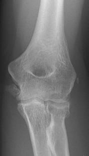
Case report: We report a case of 30-year-old female presenting with classical signs of cubital tunnel syndrome-positive Wartenberg's and Froment's signs, hypoesthesia in the fourth and fifth finger with decreased finger duction strength but without gross hypotrophy of interosseous and hypothenar muscles. Tinel's sign was positive over the ulnar sulcus and an accessory ossicle was found on the elbow radiograph within the ulnar sulcus. The first signs of calcification in this patient were reported 6 years prior in a follow-up after the dislocation of her elbow joint following a bike accident. The EMG confirmed ulnar nerve neuropathy in the elbow area. The ossicle was extirpated, the ulnar nerve was decompressed in the ulnar sulcus in a standard manner and the symptoms quickly resolved. The patient has been regularly visiting our outpatient clinic for the next 12 years without any complaints considering her elbow and the ulnar nerve.
Conclusion: This is a rare case of cubital tunnel syndrome caused by an accessory ossicle of traumatic origin. Simple bone extirpation with ulnar nerve release followed by anterior subcutaneous transposition is the recommended method of treatment. No report of congenital accessory bones causing ulnar nerve compression in the elbow exists in the literature.
期刊介绍:
Anatomy is a morphological science which cannot fail to interest the clinician. The practical application of anatomical research to clinical problems necessitates special adaptation and selectivity in choosing from numerous international works. Although there is a tendency to believe that meaningful advances in anatomy are unlikely, constant revision is necessary. Surgical and Radiologic Anatomy, the first international journal of Clinical anatomy has been created in this spirit.
Its goal is to serve clinicians, regardless of speciality-physicians, surgeons, radiologists or other specialists-as an indispensable aid with which they can improve their knowledge of anatomy. Each issue includes: Original papers, review articles, articles on the anatomical bases of medical, surgical and radiological techniques, articles of normal radiologic anatomy, brief reviews of anatomical publications of clinical interest.
Particular attention is given to high quality illustrations, which are indispensable for a better understanding of anatomical problems.
Surgical and Radiologic Anatomy is a journal written by anatomists for clinicians with a special interest in anatomy.


 分享
分享
 求助内容:
求助内容: 应助结果提醒方式:
 扫码关注我们
