{"title":"头臂动脉、右颈总动脉和右锁骨下动脉的联合鞘内路线。","authors":"Vedat Yaman, Selin Ardali Duzgun, Tuncay Hazirolan","doi":"10.1007/s00276-023-03228-2","DOIUrl":null,"url":null,"abstract":"<p><strong>Purpose: </strong>We present an extremely rare vascular variant in which the brachiocephalic artery, right common carotid artery, and right subclavian artery course through the right lobe of the thyroid gland.</p><p><strong>Methods: </strong>A 54-year-old woman underwent a coronary computed tomography (CT) angiography examination with the suspicion of infective endocarditis.</p><p><strong>Results: </strong>Unexpectedly, the distal brachiocephalic artery, the proximal right common carotid artery, and right subclavian artery had a course through the right lobe of the thyroid gland. Otherwise, the arcus aorta branching pattern was normal.</p><p><strong>Conclusion: </strong>The supraaortic major branches seldom have intrathyroidal course. The intrathyroidal course of the right common carotid artery was described previously only in one case. But, to our best knowledge, the combined intrathyroidal course of these three major vessels has not been previously reported. Although asymptomatic, such variations may complicate lower neck procedures involving thyroidectomies and thyroid biopsies if undetected and unreported. So, the awareness of this atypical course while reporting CT examinations is crucial prior to neck interventions.</p>","PeriodicalId":49296,"journal":{"name":"Surgical and Radiologic Anatomy","volume":" ","pages":"1149-1151"},"PeriodicalIF":1.2000,"publicationDate":"2023-09-01","publicationTypes":"Journal Article","fieldsOfStudy":null,"isOpenAccess":false,"openAccessPdf":"","citationCount":"0","resultStr":"{\"title\":\"The combined intrathyroidal course of the brachiocephalic artery, right common carotid artery and right subclavian artery.\",\"authors\":\"Vedat Yaman, Selin Ardali Duzgun, Tuncay Hazirolan\",\"doi\":\"10.1007/s00276-023-03228-2\",\"DOIUrl\":null,\"url\":null,\"abstract\":\"<p><strong>Purpose: </strong>We present an extremely rare vascular variant in which the brachiocephalic artery, right common carotid artery, and right subclavian artery course through the right lobe of the thyroid gland.</p><p><strong>Methods: </strong>A 54-year-old woman underwent a coronary computed tomography (CT) angiography examination with the suspicion of infective endocarditis.</p><p><strong>Results: </strong>Unexpectedly, the distal brachiocephalic artery, the proximal right common carotid artery, and right subclavian artery had a course through the right lobe of the thyroid gland. Otherwise, the arcus aorta branching pattern was normal.</p><p><strong>Conclusion: </strong>The supraaortic major branches seldom have intrathyroidal course. The intrathyroidal course of the right common carotid artery was described previously only in one case. But, to our best knowledge, the combined intrathyroidal course of these three major vessels has not been previously reported. Although asymptomatic, such variations may complicate lower neck procedures involving thyroidectomies and thyroid biopsies if undetected and unreported. So, the awareness of this atypical course while reporting CT examinations is crucial prior to neck interventions.</p>\",\"PeriodicalId\":49296,\"journal\":{\"name\":\"Surgical and Radiologic Anatomy\",\"volume\":\" \",\"pages\":\"1149-1151\"},\"PeriodicalIF\":1.2000,\"publicationDate\":\"2023-09-01\",\"publicationTypes\":\"Journal Article\",\"fieldsOfStudy\":null,\"isOpenAccess\":false,\"openAccessPdf\":\"\",\"citationCount\":\"0\",\"resultStr\":null,\"platform\":\"Semanticscholar\",\"paperid\":null,\"PeriodicalName\":\"Surgical and Radiologic Anatomy\",\"FirstCategoryId\":\"3\",\"ListUrlMain\":\"https://doi.org/10.1007/s00276-023-03228-2\",\"RegionNum\":4,\"RegionCategory\":\"医学\",\"ArticlePicture\":[],\"TitleCN\":null,\"AbstractTextCN\":null,\"PMCID\":null,\"EPubDate\":\"2023/8/15 0:00:00\",\"PubModel\":\"Epub\",\"JCR\":\"Q3\",\"JCRName\":\"ANATOMY & MORPHOLOGY\",\"Score\":null,\"Total\":0}","platform":"Semanticscholar","paperid":null,"PeriodicalName":"Surgical and Radiologic Anatomy","FirstCategoryId":"3","ListUrlMain":"https://doi.org/10.1007/s00276-023-03228-2","RegionNum":4,"RegionCategory":"医学","ArticlePicture":[],"TitleCN":null,"AbstractTextCN":null,"PMCID":null,"EPubDate":"2023/8/15 0:00:00","PubModel":"Epub","JCR":"Q3","JCRName":"ANATOMY & MORPHOLOGY","Score":null,"Total":0}
The combined intrathyroidal course of the brachiocephalic artery, right common carotid artery and right subclavian artery.
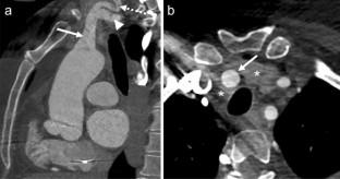
Purpose: We present an extremely rare vascular variant in which the brachiocephalic artery, right common carotid artery, and right subclavian artery course through the right lobe of the thyroid gland.
Methods: A 54-year-old woman underwent a coronary computed tomography (CT) angiography examination with the suspicion of infective endocarditis.
Results: Unexpectedly, the distal brachiocephalic artery, the proximal right common carotid artery, and right subclavian artery had a course through the right lobe of the thyroid gland. Otherwise, the arcus aorta branching pattern was normal.
Conclusion: The supraaortic major branches seldom have intrathyroidal course. The intrathyroidal course of the right common carotid artery was described previously only in one case. But, to our best knowledge, the combined intrathyroidal course of these three major vessels has not been previously reported. Although asymptomatic, such variations may complicate lower neck procedures involving thyroidectomies and thyroid biopsies if undetected and unreported. So, the awareness of this atypical course while reporting CT examinations is crucial prior to neck interventions.
期刊介绍:
Anatomy is a morphological science which cannot fail to interest the clinician. The practical application of anatomical research to clinical problems necessitates special adaptation and selectivity in choosing from numerous international works. Although there is a tendency to believe that meaningful advances in anatomy are unlikely, constant revision is necessary. Surgical and Radiologic Anatomy, the first international journal of Clinical anatomy has been created in this spirit.
Its goal is to serve clinicians, regardless of speciality-physicians, surgeons, radiologists or other specialists-as an indispensable aid with which they can improve their knowledge of anatomy. Each issue includes: Original papers, review articles, articles on the anatomical bases of medical, surgical and radiological techniques, articles of normal radiologic anatomy, brief reviews of anatomical publications of clinical interest.
Particular attention is given to high quality illustrations, which are indispensable for a better understanding of anatomical problems.
Surgical and Radiologic Anatomy is a journal written by anatomists for clinicians with a special interest in anatomy.


 分享
分享
 求助内容:
求助内容: 应助结果提醒方式:
 扫码关注我们
