{"title":"磁共振指纹技术定量测定Heschl脑回内禀T1和T2。","authors":"Sho Maruyama, Sayuri Tatsuo, Soichiro Tatsuo, Saya Iida, Fumiyasu Tsushima, Satoru Ide, Shingo Kakeda","doi":"10.2463/mrms.mp.2021-0144","DOIUrl":null,"url":null,"abstract":"<p><strong>Purpose: </strong>The human primary auditory cortex is located in the Heschl's gyrus (HG). To assess the intrinsic MR property in the gray matter of the HG (GM-HG) with T1 and T2 values using a commercially available MR fingerprinting (MRF) technique.</p><p><strong>Methods: </strong>The subjects were 10 healthy volunteers (with 20 HGs; mean age, 31.5 years old; range, 25-53 years old). Coronal T1 and T2 maps were obtained with commercially available MRF using a 3-Tesla MR system. Two radiologists measured the T1 and T2 values of the GM-HG, the GM in the superior temporal gyrus (GM-STG), and the GM in the middle temporal gyrus (GM-MTG) by drawing a ROI on coronal maps.</p><p><strong>Results: </strong>For both radiologists, the mean T1 and T2 values of the GM-HG were significantly lower than those in the GM-STG or GM-MTG (P < 0.01). The interobserver reliability using the intraclass correlation coefficients (ICC) (2,1) showed strong agreement for the measurement of the T1 and T2 values (ICCs =⃥ 0.80 and 0.78 for T1 and T2 values, respectively).</p><p><strong>Conclusion: </strong>The T1 and T2 values on MRF for the GM-HG were lower than those for the GM-STG and GM-MTG, likely reflecting a higher myelin content and iron deposition in the GM-HG. Quantitative measurements using the MRF can clarify cortical properties with high reliability, which may indicate that MRF mapping provides new insights into the structure of the human cortical GM.</p>","PeriodicalId":18119,"journal":{"name":"Magnetic Resonance in Medical Sciences","volume":"22 1","pages":"95-101"},"PeriodicalIF":2.5000,"publicationDate":"2023-01-01","publicationTypes":"Journal Article","fieldsOfStudy":null,"isOpenAccess":false,"openAccessPdf":"https://ftp.ncbi.nlm.nih.gov/pub/pmc/oa_pdf/56/72/mrms-22-95.PMC9849413.pdf","citationCount":"0","resultStr":"{\"title\":\"Quantification of the Intrinsic T1 and T2 of Heschl's Gyri with MR Fingerprinting.\",\"authors\":\"Sho Maruyama, Sayuri Tatsuo, Soichiro Tatsuo, Saya Iida, Fumiyasu Tsushima, Satoru Ide, Shingo Kakeda\",\"doi\":\"10.2463/mrms.mp.2021-0144\",\"DOIUrl\":null,\"url\":null,\"abstract\":\"<p><strong>Purpose: </strong>The human primary auditory cortex is located in the Heschl's gyrus (HG). To assess the intrinsic MR property in the gray matter of the HG (GM-HG) with T1 and T2 values using a commercially available MR fingerprinting (MRF) technique.</p><p><strong>Methods: </strong>The subjects were 10 healthy volunteers (with 20 HGs; mean age, 31.5 years old; range, 25-53 years old). Coronal T1 and T2 maps were obtained with commercially available MRF using a 3-Tesla MR system. Two radiologists measured the T1 and T2 values of the GM-HG, the GM in the superior temporal gyrus (GM-STG), and the GM in the middle temporal gyrus (GM-MTG) by drawing a ROI on coronal maps.</p><p><strong>Results: </strong>For both radiologists, the mean T1 and T2 values of the GM-HG were significantly lower than those in the GM-STG or GM-MTG (P < 0.01). The interobserver reliability using the intraclass correlation coefficients (ICC) (2,1) showed strong agreement for the measurement of the T1 and T2 values (ICCs =⃥ 0.80 and 0.78 for T1 and T2 values, respectively).</p><p><strong>Conclusion: </strong>The T1 and T2 values on MRF for the GM-HG were lower than those for the GM-STG and GM-MTG, likely reflecting a higher myelin content and iron deposition in the GM-HG. Quantitative measurements using the MRF can clarify cortical properties with high reliability, which may indicate that MRF mapping provides new insights into the structure of the human cortical GM.</p>\",\"PeriodicalId\":18119,\"journal\":{\"name\":\"Magnetic Resonance in Medical Sciences\",\"volume\":\"22 1\",\"pages\":\"95-101\"},\"PeriodicalIF\":2.5000,\"publicationDate\":\"2023-01-01\",\"publicationTypes\":\"Journal Article\",\"fieldsOfStudy\":null,\"isOpenAccess\":false,\"openAccessPdf\":\"https://ftp.ncbi.nlm.nih.gov/pub/pmc/oa_pdf/56/72/mrms-22-95.PMC9849413.pdf\",\"citationCount\":\"0\",\"resultStr\":null,\"platform\":\"Semanticscholar\",\"paperid\":null,\"PeriodicalName\":\"Magnetic Resonance in Medical Sciences\",\"FirstCategoryId\":\"3\",\"ListUrlMain\":\"https://doi.org/10.2463/mrms.mp.2021-0144\",\"RegionNum\":3,\"RegionCategory\":\"医学\",\"ArticlePicture\":[],\"TitleCN\":null,\"AbstractTextCN\":null,\"PMCID\":null,\"EPubDate\":\"\",\"PubModel\":\"\",\"JCR\":\"Q2\",\"JCRName\":\"RADIOLOGY, NUCLEAR MEDICINE & MEDICAL IMAGING\",\"Score\":null,\"Total\":0}","platform":"Semanticscholar","paperid":null,"PeriodicalName":"Magnetic Resonance in Medical Sciences","FirstCategoryId":"3","ListUrlMain":"https://doi.org/10.2463/mrms.mp.2021-0144","RegionNum":3,"RegionCategory":"医学","ArticlePicture":[],"TitleCN":null,"AbstractTextCN":null,"PMCID":null,"EPubDate":"","PubModel":"","JCR":"Q2","JCRName":"RADIOLOGY, NUCLEAR MEDICINE & MEDICAL IMAGING","Score":null,"Total":0}
Quantification of the Intrinsic T1 and T2 of Heschl's Gyri with MR Fingerprinting.
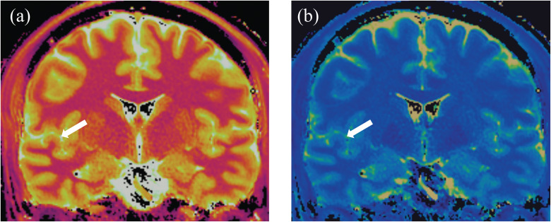
Purpose: The human primary auditory cortex is located in the Heschl's gyrus (HG). To assess the intrinsic MR property in the gray matter of the HG (GM-HG) with T1 and T2 values using a commercially available MR fingerprinting (MRF) technique.
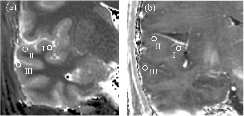
Methods: The subjects were 10 healthy volunteers (with 20 HGs; mean age, 31.5 years old; range, 25-53 years old). Coronal T1 and T2 maps were obtained with commercially available MRF using a 3-Tesla MR system. Two radiologists measured the T1 and T2 values of the GM-HG, the GM in the superior temporal gyrus (GM-STG), and the GM in the middle temporal gyrus (GM-MTG) by drawing a ROI on coronal maps.
Results: For both radiologists, the mean T1 and T2 values of the GM-HG were significantly lower than those in the GM-STG or GM-MTG (P < 0.01). The interobserver reliability using the intraclass correlation coefficients (ICC) (2,1) showed strong agreement for the measurement of the T1 and T2 values (ICCs =⃥ 0.80 and 0.78 for T1 and T2 values, respectively).
Conclusion: The T1 and T2 values on MRF for the GM-HG were lower than those for the GM-STG and GM-MTG, likely reflecting a higher myelin content and iron deposition in the GM-HG. Quantitative measurements using the MRF can clarify cortical properties with high reliability, which may indicate that MRF mapping provides new insights into the structure of the human cortical GM.
期刊介绍:
Magnetic Resonance in Medical Sciences (MRMS or Magn
Reson Med Sci) is an international journal pursuing the
publication of original articles contributing to the progress
of magnetic resonance in the field of biomedical sciences
including technical developments and clinical applications.
MRMS is an official journal of the Japanese Society for
Magnetic Resonance in Medicine (JSMRM).


 分享
分享
 求助内容:
求助内容: 应助结果提醒方式:
 扫码关注我们
