{"title":"太平洋蓝鳍金枪鱼椎体生长过程中的侧骨脊扩张和内部组织置换","authors":"Misaki Sakashita, Shigeru Kondo, Naoyuki Wada","doi":"10.1002/jmor.21666","DOIUrl":null,"url":null,"abstract":"<p>Vertebral growth is an essential developmental process to support the expansion of the vertebrate body. In teleosts, the lateral side of the vertebral bodies develops to form different structures among species in the late stages of vertebral growth, although lateral structures are not apparent in the early stages. Lateral structures are one of the structural features that determine the diversity of teleost vertebrae. However, explanations for the formation of lateral structures are conflicting because few reports have investigated the growth of teleost vertebral bodies. To clarify the growth process, we analyzed the morphological changes in the vertebral body of Pacific bluefin tuna <i>Thunnus orientalis</i> at different developmental stages using micro-computed tomography (CT) scans. The micro-CT scans showed that the vertebral centrum formed a plate-like ridge on the lateral side along the cranial–caudal direction and extended laterally with increasing thickness. Simultaneously, the proximal region of the lateral ridges became porous as the vertebrae grew to form bone marrow cavities. Furthermore, we used histological observations to describe the relationship between these morphological changes and osteoblast and osteoclast activities. Osteoblasts accumulated on the distal edges of the lateral ridges, whereas osteoclasts were distributed in the bone marrow cavities. These observations suggest that bone resorption occurs proximally to form bone marrow cavities in addition to bone synthesis at the edges of the lateral ridges. The bone marrow cavities were occupied by blood vessels, extracellular matrix, and adipocytes, and the internal tissue composition changed to increase the area of adipose tissue. Because the ratio of bone volume decreases in large vertebrae, bone formation and resorption are regulated to separate the external cortical and internal trabecular bones to support the vertebrae. This study is the first to report the formation of lateral structures and can be applied to similar lateral structures in the vertebrae of other teleost species.</p>","PeriodicalId":16528,"journal":{"name":"Journal of Morphology","volume":"285 2","pages":""},"PeriodicalIF":1.4000,"publicationDate":"2023-12-21","publicationTypes":"Journal Article","fieldsOfStudy":null,"isOpenAccess":false,"openAccessPdf":"https://onlinelibrary.wiley.com/doi/epdf/10.1002/jmor.21666","citationCount":"0","resultStr":"{\"title\":\"Lateral bone ridge expansion and internal tissue replacement for vertebral body growth in Pacific bluefin tuna Thunnus orientalis\",\"authors\":\"Misaki Sakashita, Shigeru Kondo, Naoyuki Wada\",\"doi\":\"10.1002/jmor.21666\",\"DOIUrl\":null,\"url\":null,\"abstract\":\"<p>Vertebral growth is an essential developmental process to support the expansion of the vertebrate body. In teleosts, the lateral side of the vertebral bodies develops to form different structures among species in the late stages of vertebral growth, although lateral structures are not apparent in the early stages. Lateral structures are one of the structural features that determine the diversity of teleost vertebrae. However, explanations for the formation of lateral structures are conflicting because few reports have investigated the growth of teleost vertebral bodies. To clarify the growth process, we analyzed the morphological changes in the vertebral body of Pacific bluefin tuna <i>Thunnus orientalis</i> at different developmental stages using micro-computed tomography (CT) scans. The micro-CT scans showed that the vertebral centrum formed a plate-like ridge on the lateral side along the cranial–caudal direction and extended laterally with increasing thickness. Simultaneously, the proximal region of the lateral ridges became porous as the vertebrae grew to form bone marrow cavities. Furthermore, we used histological observations to describe the relationship between these morphological changes and osteoblast and osteoclast activities. Osteoblasts accumulated on the distal edges of the lateral ridges, whereas osteoclasts were distributed in the bone marrow cavities. These observations suggest that bone resorption occurs proximally to form bone marrow cavities in addition to bone synthesis at the edges of the lateral ridges. The bone marrow cavities were occupied by blood vessels, extracellular matrix, and adipocytes, and the internal tissue composition changed to increase the area of adipose tissue. Because the ratio of bone volume decreases in large vertebrae, bone formation and resorption are regulated to separate the external cortical and internal trabecular bones to support the vertebrae. This study is the first to report the formation of lateral structures and can be applied to similar lateral structures in the vertebrae of other teleost species.</p>\",\"PeriodicalId\":16528,\"journal\":{\"name\":\"Journal of Morphology\",\"volume\":\"285 2\",\"pages\":\"\"},\"PeriodicalIF\":1.4000,\"publicationDate\":\"2023-12-21\",\"publicationTypes\":\"Journal Article\",\"fieldsOfStudy\":null,\"isOpenAccess\":false,\"openAccessPdf\":\"https://onlinelibrary.wiley.com/doi/epdf/10.1002/jmor.21666\",\"citationCount\":\"0\",\"resultStr\":null,\"platform\":\"Semanticscholar\",\"paperid\":null,\"PeriodicalName\":\"Journal of Morphology\",\"FirstCategoryId\":\"3\",\"ListUrlMain\":\"https://onlinelibrary.wiley.com/doi/10.1002/jmor.21666\",\"RegionNum\":4,\"RegionCategory\":\"医学\",\"ArticlePicture\":[],\"TitleCN\":null,\"AbstractTextCN\":null,\"PMCID\":null,\"EPubDate\":\"\",\"PubModel\":\"\",\"JCR\":\"Q2\",\"JCRName\":\"ANATOMY & MORPHOLOGY\",\"Score\":null,\"Total\":0}","platform":"Semanticscholar","paperid":null,"PeriodicalName":"Journal of Morphology","FirstCategoryId":"3","ListUrlMain":"https://onlinelibrary.wiley.com/doi/10.1002/jmor.21666","RegionNum":4,"RegionCategory":"医学","ArticlePicture":[],"TitleCN":null,"AbstractTextCN":null,"PMCID":null,"EPubDate":"","PubModel":"","JCR":"Q2","JCRName":"ANATOMY & MORPHOLOGY","Score":null,"Total":0}
Lateral bone ridge expansion and internal tissue replacement for vertebral body growth in Pacific bluefin tuna Thunnus orientalis
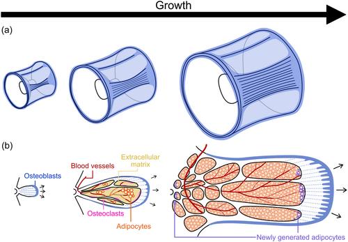
Vertebral growth is an essential developmental process to support the expansion of the vertebrate body. In teleosts, the lateral side of the vertebral bodies develops to form different structures among species in the late stages of vertebral growth, although lateral structures are not apparent in the early stages. Lateral structures are one of the structural features that determine the diversity of teleost vertebrae. However, explanations for the formation of lateral structures are conflicting because few reports have investigated the growth of teleost vertebral bodies. To clarify the growth process, we analyzed the morphological changes in the vertebral body of Pacific bluefin tuna Thunnus orientalis at different developmental stages using micro-computed tomography (CT) scans. The micro-CT scans showed that the vertebral centrum formed a plate-like ridge on the lateral side along the cranial–caudal direction and extended laterally with increasing thickness. Simultaneously, the proximal region of the lateral ridges became porous as the vertebrae grew to form bone marrow cavities. Furthermore, we used histological observations to describe the relationship between these morphological changes and osteoblast and osteoclast activities. Osteoblasts accumulated on the distal edges of the lateral ridges, whereas osteoclasts were distributed in the bone marrow cavities. These observations suggest that bone resorption occurs proximally to form bone marrow cavities in addition to bone synthesis at the edges of the lateral ridges. The bone marrow cavities were occupied by blood vessels, extracellular matrix, and adipocytes, and the internal tissue composition changed to increase the area of adipose tissue. Because the ratio of bone volume decreases in large vertebrae, bone formation and resorption are regulated to separate the external cortical and internal trabecular bones to support the vertebrae. This study is the first to report the formation of lateral structures and can be applied to similar lateral structures in the vertebrae of other teleost species.
期刊介绍:
The Journal of Morphology welcomes articles of original research in cytology, protozoology, embryology, and general morphology. Articles generally should not exceed 35 printed pages. Preliminary notices or articles of a purely descriptive morphological or taxonomic nature are not included. No paper which has already been published will be accepted, nor will simultaneous publications elsewhere be allowed.
The Journal of Morphology publishes research in functional, comparative, evolutionary and developmental morphology from vertebrates and invertebrates. Human and veterinary anatomy or paleontology are considered when an explicit connection to neontological animal morphology is presented, and the paper contains relevant information for the community of animal morphologists. Based on our long tradition, we continue to seek publishing the best papers in animal morphology.


 分享
分享
 求助内容:
求助内容: 应助结果提醒方式:
 扫码关注我们
