{"title":"弥漫性大 B 细胞淋巴瘤(非特异性)中淋巴瘤细胞和肿瘤免疫细胞上的程序性细胞死亡蛋白 1 及其配体的预后意义。","authors":"Teja Cas Slak, Simona Miceska, Gorana Gasljevic, Lucka Boltezar, Veronika Kloboves-Prevodnik","doi":"10.2478/raon-2024-0010","DOIUrl":null,"url":null,"abstract":"<p><strong>Background: </strong>Diffuse large B-cell lymphoma, not otherwise specified (DLBCL, NOS) is the most common type non-Hodgkin's lymphoma, where the treatment of relapsed/refractory cases is the major challenge. Programmed cell death protein 1 (PD-1) and its ligand PD-L1 play a crucial role in the negative regulation of the immune response against the disease. The aim of the study was to analyze the expression of PD-1 and PD-L1 on lymphoma cells (LCs) and tumor-immune cells (TICs) and to investigate their correlation with outcome.</p><p><strong>Patients and methods: </strong>Samples from 283 patients diagnosed with DLBCL, NOS (both germinal center B cell like [GCB] and non-GCB subtypes) were included in the study. Expression of PD-1 and PD-L1 was determined using double immunohistochemical staining (D-IHC) for PD-1/PAX5 and PD-L1/PAX5 on tissue microarrays. LCs were highlighted by D-IHC to obtain more accurate results. Clinical data and histologic diagnoses were obtained from electronic data records. We correlated clinical characteristics, and PD-1 and PD-L1 expression on LCs and TICs with progression-free survival (PFS) and overall survival (OS).</p><p><strong>Results: </strong>Expression of PD-1 on TICs was observed in 38.4% and on LCs in 8.8% of cases, while PD-L1 was expressed on TICs in 46.8% and on LCs in 6.5% of cases. PD-L1 expression on LCs was more frequent in non-GCB subtype (p = 0.047). In addition, patients with PD-L1 expression on LCs had significantly shorter PFS (p = 0.015), and the expression retained significant in the multivariate model (p = 0.034).</p><p><strong>Conclusions: </strong>PD-L1 was more frequently expressed in LCs of the non-GCB subtype. Additionally, PD-L1 in LCs may predict shorter PFS time. D-IHC staining for PD-L1/PAX5 is a feasible method to assess PD-L1 expression on LCs of DLBCL, NOS patients and can be used to identify patients who may benefit from targeted immunotherapy with checkpoint inhibitors.</p>","PeriodicalId":21034,"journal":{"name":"Radiology and Oncology","volume":"58 1","pages":"99-109"},"PeriodicalIF":2.2000,"publicationDate":"2024-02-21","publicationTypes":"Journal Article","fieldsOfStudy":null,"isOpenAccess":false,"openAccessPdf":"https://www.ncbi.nlm.nih.gov/pmc/articles/PMC10878775/pdf/","citationCount":"0","resultStr":"{\"title\":\"The prognostic significance of programmed cell death protein 1 and its ligand on lymphoma cells and tumor-immune cells in diffuse large B-cell lymphoma, not otherwise specified.\",\"authors\":\"Teja Cas Slak, Simona Miceska, Gorana Gasljevic, Lucka Boltezar, Veronika Kloboves-Prevodnik\",\"doi\":\"10.2478/raon-2024-0010\",\"DOIUrl\":null,\"url\":null,\"abstract\":\"<p><strong>Background: </strong>Diffuse large B-cell lymphoma, not otherwise specified (DLBCL, NOS) is the most common type non-Hodgkin's lymphoma, where the treatment of relapsed/refractory cases is the major challenge. Programmed cell death protein 1 (PD-1) and its ligand PD-L1 play a crucial role in the negative regulation of the immune response against the disease. The aim of the study was to analyze the expression of PD-1 and PD-L1 on lymphoma cells (LCs) and tumor-immune cells (TICs) and to investigate their correlation with outcome.</p><p><strong>Patients and methods: </strong>Samples from 283 patients diagnosed with DLBCL, NOS (both germinal center B cell like [GCB] and non-GCB subtypes) were included in the study. Expression of PD-1 and PD-L1 was determined using double immunohistochemical staining (D-IHC) for PD-1/PAX5 and PD-L1/PAX5 on tissue microarrays. LCs were highlighted by D-IHC to obtain more accurate results. Clinical data and histologic diagnoses were obtained from electronic data records. We correlated clinical characteristics, and PD-1 and PD-L1 expression on LCs and TICs with progression-free survival (PFS) and overall survival (OS).</p><p><strong>Results: </strong>Expression of PD-1 on TICs was observed in 38.4% and on LCs in 8.8% of cases, while PD-L1 was expressed on TICs in 46.8% and on LCs in 6.5% of cases. PD-L1 expression on LCs was more frequent in non-GCB subtype (p = 0.047). In addition, patients with PD-L1 expression on LCs had significantly shorter PFS (p = 0.015), and the expression retained significant in the multivariate model (p = 0.034).</p><p><strong>Conclusions: </strong>PD-L1 was more frequently expressed in LCs of the non-GCB subtype. Additionally, PD-L1 in LCs may predict shorter PFS time. D-IHC staining for PD-L1/PAX5 is a feasible method to assess PD-L1 expression on LCs of DLBCL, NOS patients and can be used to identify patients who may benefit from targeted immunotherapy with checkpoint inhibitors.</p>\",\"PeriodicalId\":21034,\"journal\":{\"name\":\"Radiology and Oncology\",\"volume\":\"58 1\",\"pages\":\"99-109\"},\"PeriodicalIF\":2.2000,\"publicationDate\":\"2024-02-21\",\"publicationTypes\":\"Journal Article\",\"fieldsOfStudy\":null,\"isOpenAccess\":false,\"openAccessPdf\":\"https://www.ncbi.nlm.nih.gov/pmc/articles/PMC10878775/pdf/\",\"citationCount\":\"0\",\"resultStr\":null,\"platform\":\"Semanticscholar\",\"paperid\":null,\"PeriodicalName\":\"Radiology and Oncology\",\"FirstCategoryId\":\"3\",\"ListUrlMain\":\"https://doi.org/10.2478/raon-2024-0010\",\"RegionNum\":4,\"RegionCategory\":\"医学\",\"ArticlePicture\":[],\"TitleCN\":null,\"AbstractTextCN\":null,\"PMCID\":null,\"EPubDate\":\"2024/3/1 0:00:00\",\"PubModel\":\"eCollection\",\"JCR\":\"Q3\",\"JCRName\":\"ONCOLOGY\",\"Score\":null,\"Total\":0}","platform":"Semanticscholar","paperid":null,"PeriodicalName":"Radiology and Oncology","FirstCategoryId":"3","ListUrlMain":"https://doi.org/10.2478/raon-2024-0010","RegionNum":4,"RegionCategory":"医学","ArticlePicture":[],"TitleCN":null,"AbstractTextCN":null,"PMCID":null,"EPubDate":"2024/3/1 0:00:00","PubModel":"eCollection","JCR":"Q3","JCRName":"ONCOLOGY","Score":null,"Total":0}
The prognostic significance of programmed cell death protein 1 and its ligand on lymphoma cells and tumor-immune cells in diffuse large B-cell lymphoma, not otherwise specified.
Background: Diffuse large B-cell lymphoma, not otherwise specified (DLBCL, NOS) is the most common type non-Hodgkin's lymphoma, where the treatment of relapsed/refractory cases is the major challenge. Programmed cell death protein 1 (PD-1) and its ligand PD-L1 play a crucial role in the negative regulation of the immune response against the disease. The aim of the study was to analyze the expression of PD-1 and PD-L1 on lymphoma cells (LCs) and tumor-immune cells (TICs) and to investigate their correlation with outcome.
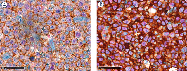
Patients and methods: Samples from 283 patients diagnosed with DLBCL, NOS (both germinal center B cell like [GCB] and non-GCB subtypes) were included in the study. Expression of PD-1 and PD-L1 was determined using double immunohistochemical staining (D-IHC) for PD-1/PAX5 and PD-L1/PAX5 on tissue microarrays. LCs were highlighted by D-IHC to obtain more accurate results. Clinical data and histologic diagnoses were obtained from electronic data records. We correlated clinical characteristics, and PD-1 and PD-L1 expression on LCs and TICs with progression-free survival (PFS) and overall survival (OS).
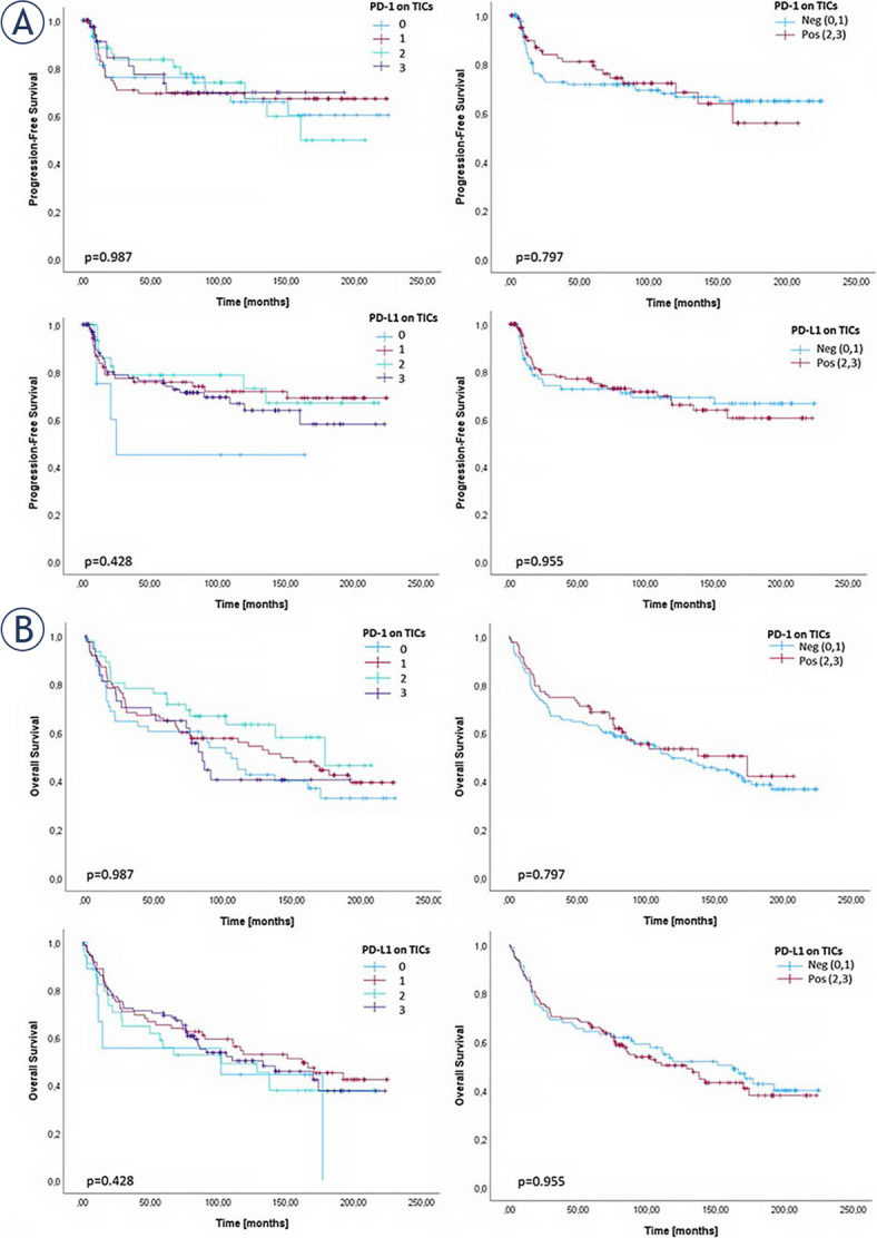
Results: Expression of PD-1 on TICs was observed in 38.4% and on LCs in 8.8% of cases, while PD-L1 was expressed on TICs in 46.8% and on LCs in 6.5% of cases. PD-L1 expression on LCs was more frequent in non-GCB subtype (p = 0.047). In addition, patients with PD-L1 expression on LCs had significantly shorter PFS (p = 0.015), and the expression retained significant in the multivariate model (p = 0.034).
Conclusions: PD-L1 was more frequently expressed in LCs of the non-GCB subtype. Additionally, PD-L1 in LCs may predict shorter PFS time. D-IHC staining for PD-L1/PAX5 is a feasible method to assess PD-L1 expression on LCs of DLBCL, NOS patients and can be used to identify patients who may benefit from targeted immunotherapy with checkpoint inhibitors.
期刊介绍:
Radiology and Oncology is a multidisciplinary journal devoted to the publishing original and high quality scientific papers and review articles, pertinent to diagnostic and interventional radiology, computerized tomography, magnetic resonance, ultrasound, nuclear medicine, radiotherapy, clinical and experimental oncology, radiobiology, medical physics and radiation protection. Therefore, the scope of the journal is to cover beside radiology the diagnostic and therapeutic aspects in oncology, which distinguishes it from other journals in the field.


 分享
分享
 求助内容:
求助内容: 应助结果提醒方式:
 扫码关注我们
