Roshni Ramesh, Anoop Sasi, Shahana C Mohamed, Sonia P Joseph
{"title":"\"压迫坏死\"--种植体早期失败的原因之一?病例报告和文献综述。","authors":"Roshni Ramesh, Anoop Sasi, Shahana C Mohamed, Sonia P Joseph","doi":"10.2147/CCIDE.S453798","DOIUrl":null,"url":null,"abstract":"<p><strong>Purpose: </strong>Compression necrosis refers to bone tissue damage that can occur when excessive pressure or force is applied to surrounding bone during implant placement. This pressure can compromise blood supply to the bone, leading to necrosis. Compression necrosis is a concern, because it can affect the stability and long-term success of dental implant.</p><p><strong>Patients and methods: </strong>This case report highlights a case of early bone loss and implant failure possibly due to compression necrosis. Clinical data, photographs, radiographs, blood examination report and histology were presented to document the early failure of an implant placed in the mandibular left posterior region of a 33-year-old female patient.</p><p><strong>Results: </strong>Radiograph taken six weeks after implant placement showed severe angular defect. Therefore, the implant was surgically removed. Histological examination of the area showed bony trabeculae with an absence of osteoblastic riming, suggestive of necrotic bone.</p><p><strong>Conclusion: </strong>Using excessive torque values when placing implants in dense bones can increase the risk of implant failure due to bone over compression. Dental professionals must follow the manufacturer's instructions and employ quality surgical techniques during implant placement into dense cortical bone to minimise risks.</p>","PeriodicalId":10445,"journal":{"name":"Clinical, Cosmetic and Investigational Dentistry","volume":"16 ","pages":"43-52"},"PeriodicalIF":1.8000,"publicationDate":"2024-03-07","publicationTypes":"Journal Article","fieldsOfStudy":null,"isOpenAccess":false,"openAccessPdf":"https://www.ncbi.nlm.nih.gov/pmc/articles/PMC10926919/pdf/","citationCount":"0","resultStr":"{\"title\":\"\\\"Compression Necrosis\\\" - A Cause of Concern for Early Implant Failure? Case Report and Review of Literature.\",\"authors\":\"Roshni Ramesh, Anoop Sasi, Shahana C Mohamed, Sonia P Joseph\",\"doi\":\"10.2147/CCIDE.S453798\",\"DOIUrl\":null,\"url\":null,\"abstract\":\"<p><strong>Purpose: </strong>Compression necrosis refers to bone tissue damage that can occur when excessive pressure or force is applied to surrounding bone during implant placement. This pressure can compromise blood supply to the bone, leading to necrosis. Compression necrosis is a concern, because it can affect the stability and long-term success of dental implant.</p><p><strong>Patients and methods: </strong>This case report highlights a case of early bone loss and implant failure possibly due to compression necrosis. Clinical data, photographs, radiographs, blood examination report and histology were presented to document the early failure of an implant placed in the mandibular left posterior region of a 33-year-old female patient.</p><p><strong>Results: </strong>Radiograph taken six weeks after implant placement showed severe angular defect. Therefore, the implant was surgically removed. Histological examination of the area showed bony trabeculae with an absence of osteoblastic riming, suggestive of necrotic bone.</p><p><strong>Conclusion: </strong>Using excessive torque values when placing implants in dense bones can increase the risk of implant failure due to bone over compression. Dental professionals must follow the manufacturer's instructions and employ quality surgical techniques during implant placement into dense cortical bone to minimise risks.</p>\",\"PeriodicalId\":10445,\"journal\":{\"name\":\"Clinical, Cosmetic and Investigational Dentistry\",\"volume\":\"16 \",\"pages\":\"43-52\"},\"PeriodicalIF\":1.8000,\"publicationDate\":\"2024-03-07\",\"publicationTypes\":\"Journal Article\",\"fieldsOfStudy\":null,\"isOpenAccess\":false,\"openAccessPdf\":\"https://www.ncbi.nlm.nih.gov/pmc/articles/PMC10926919/pdf/\",\"citationCount\":\"0\",\"resultStr\":null,\"platform\":\"Semanticscholar\",\"paperid\":null,\"PeriodicalName\":\"Clinical, Cosmetic and Investigational Dentistry\",\"FirstCategoryId\":\"1085\",\"ListUrlMain\":\"https://doi.org/10.2147/CCIDE.S453798\",\"RegionNum\":0,\"RegionCategory\":null,\"ArticlePicture\":[],\"TitleCN\":null,\"AbstractTextCN\":null,\"PMCID\":null,\"EPubDate\":\"2024/1/1 0:00:00\",\"PubModel\":\"eCollection\",\"JCR\":\"Q3\",\"JCRName\":\"DENTISTRY, ORAL SURGERY & MEDICINE\",\"Score\":null,\"Total\":0}","platform":"Semanticscholar","paperid":null,"PeriodicalName":"Clinical, Cosmetic and Investigational Dentistry","FirstCategoryId":"1085","ListUrlMain":"https://doi.org/10.2147/CCIDE.S453798","RegionNum":0,"RegionCategory":null,"ArticlePicture":[],"TitleCN":null,"AbstractTextCN":null,"PMCID":null,"EPubDate":"2024/1/1 0:00:00","PubModel":"eCollection","JCR":"Q3","JCRName":"DENTISTRY, ORAL SURGERY & MEDICINE","Score":null,"Total":0}
引用次数: 0
摘要
目的:压迫性坏死是指在植入种植体时,如果周围的骨受到过大的压力或力量,就会造成骨组织损伤。这种压力会损害骨的血液供应,从而导致坏死。压迫性坏死是一个令人担忧的问题,因为它会影响种植牙的稳定性和长期成功:本病例报告重点介绍了一例可能因压迫性坏死导致的早期骨质流失和种植失败的病例。通过临床数据、照片、X 光片、血液检查报告和组织学检查,记录了一名 33 岁女性患者下颌左后区种植体的早期失败:结果:种植体植入六周后拍摄的 X 光片显示存在严重的角度缺损。因此,通过手术取出了种植体。该区域的组织学检查显示有骨小梁,但无成骨细胞边缘,提示为坏死骨:结论:在致密骨中植入种植体时使用过大的扭矩值会增加因骨过度压缩而导致种植体失败的风险。牙科专业人员在将种植体植入致密皮质骨时,必须遵循制造商的说明,并采用优质的外科技术,以最大限度地降低风险。
"Compression Necrosis" - A Cause of Concern for Early Implant Failure? Case Report and Review of Literature.
Purpose: Compression necrosis refers to bone tissue damage that can occur when excessive pressure or force is applied to surrounding bone during implant placement. This pressure can compromise blood supply to the bone, leading to necrosis. Compression necrosis is a concern, because it can affect the stability and long-term success of dental implant.
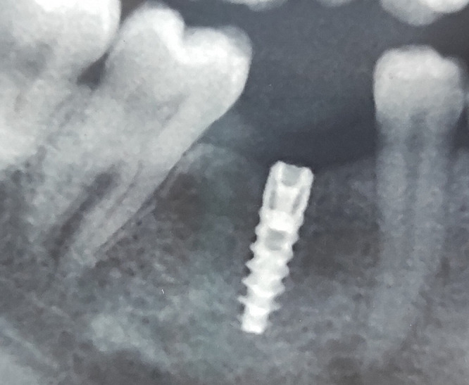
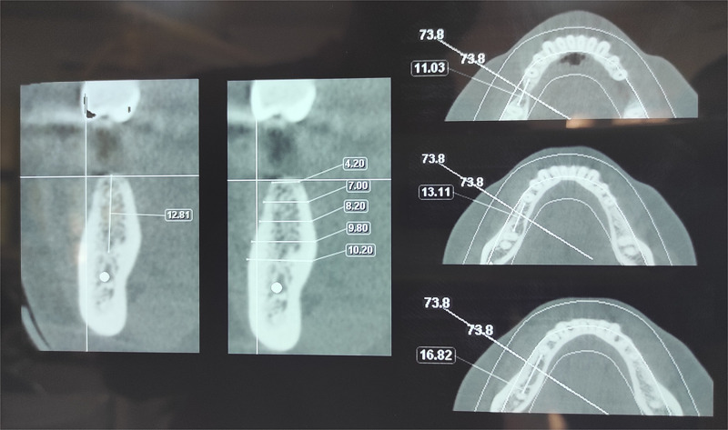
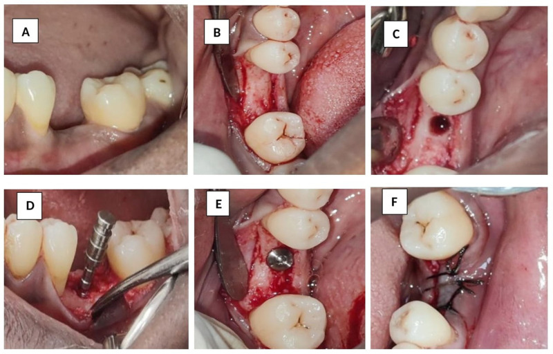
Patients and methods: This case report highlights a case of early bone loss and implant failure possibly due to compression necrosis. Clinical data, photographs, radiographs, blood examination report and histology were presented to document the early failure of an implant placed in the mandibular left posterior region of a 33-year-old female patient.
Results: Radiograph taken six weeks after implant placement showed severe angular defect. Therefore, the implant was surgically removed. Histological examination of the area showed bony trabeculae with an absence of osteoblastic riming, suggestive of necrotic bone.
Conclusion: Using excessive torque values when placing implants in dense bones can increase the risk of implant failure due to bone over compression. Dental professionals must follow the manufacturer's instructions and employ quality surgical techniques during implant placement into dense cortical bone to minimise risks.


 分享
分享
 求助内容:
求助内容: 应助结果提醒方式:
 扫码关注我们
