Bibi S van Thiel, Martine de Boer, Yanto Ridwan, Marion G J de Kleijnen, Nicole van Vliet, Janette van der Linden, Isa de Beer, Paula M van Heijningen, Wilbert P Vermeij, Jan H J Hoeijmakers, A H Jan Danser, Roland Kanaar, Dirk J Duncker, Ingrid van der Pluijm, Jeroen Essers
{"title":"混合分子和功能显微 CT 成像揭示了嗜雌性 Ercc1 小鼠心肌凋亡增加导致心力衰竭的前兆","authors":"Bibi S van Thiel, Martine de Boer, Yanto Ridwan, Marion G J de Kleijnen, Nicole van Vliet, Janette van der Linden, Isa de Beer, Paula M van Heijningen, Wilbert P Vermeij, Jan H J Hoeijmakers, A H Jan Danser, Roland Kanaar, Dirk J Duncker, Ingrid van der Pluijm, Jeroen Essers","doi":"10.1007/s11307-024-01902-4","DOIUrl":null,"url":null,"abstract":"<p><strong>Purpose: </strong>In this study, we explored the role of apoptosis as a potential biomarker for cardiac failure using functional micro-CT and fluorescence molecular tomography (FMT) imaging techniques in Ercc1 mutant mice. Ercc1 is involved in multiple DNA repair pathways, and its mutations contribute to accelerated aging phenotypes in both humans and mice, due to the accumulation of DNA lesions that impair vital DNA functions. We previously found that systemic mutations and cardiomyocyte-restricted deletion of Ercc1 in mice results in left ventricular (LV) dysfunction at older age.</p><p><strong>Procedures and results: </strong>Here we report that combined functional micro-CT and FMT imaging allowed us to detect apoptosis in systemic Ercc1 mutant mice prior to the development of overt LV dysfunction, suggesting its potential as an early indicator and contributing factor of cardiac impairment. The detection of apoptosis in vivo was feasible as early as 12 weeks of age, even when global LV function appeared normal, underscoring the potential of apoptosis as an early predictor of LV dysfunction, which subsequently manifested at 24 weeks.</p><p><strong>Conclusions: </strong>This study highlights the utility of combined functional micro-CT and FMT imaging in assessing cardiac function and detecting apoptosis, providing valuable insights into the potential of apoptosis as an early biomarker for cardiac failure.</p>","PeriodicalId":18760,"journal":{"name":"Molecular Imaging and Biology","volume":" ","pages":"628-637"},"PeriodicalIF":2.5000,"publicationDate":"2024-08-01","publicationTypes":"Journal Article","fieldsOfStudy":null,"isOpenAccess":false,"openAccessPdf":"https://www.ncbi.nlm.nih.gov/pmc/articles/PMC11281969/pdf/","citationCount":"0","resultStr":"{\"title\":\"Hybrid Molecular and Functional Micro-CT Imaging Reveals Increased Myocardial Apoptosis Preceding Cardiac Failure in Progeroid Ercc1 Mice.\",\"authors\":\"Bibi S van Thiel, Martine de Boer, Yanto Ridwan, Marion G J de Kleijnen, Nicole van Vliet, Janette van der Linden, Isa de Beer, Paula M van Heijningen, Wilbert P Vermeij, Jan H J Hoeijmakers, A H Jan Danser, Roland Kanaar, Dirk J Duncker, Ingrid van der Pluijm, Jeroen Essers\",\"doi\":\"10.1007/s11307-024-01902-4\",\"DOIUrl\":null,\"url\":null,\"abstract\":\"<p><strong>Purpose: </strong>In this study, we explored the role of apoptosis as a potential biomarker for cardiac failure using functional micro-CT and fluorescence molecular tomography (FMT) imaging techniques in Ercc1 mutant mice. Ercc1 is involved in multiple DNA repair pathways, and its mutations contribute to accelerated aging phenotypes in both humans and mice, due to the accumulation of DNA lesions that impair vital DNA functions. We previously found that systemic mutations and cardiomyocyte-restricted deletion of Ercc1 in mice results in left ventricular (LV) dysfunction at older age.</p><p><strong>Procedures and results: </strong>Here we report that combined functional micro-CT and FMT imaging allowed us to detect apoptosis in systemic Ercc1 mutant mice prior to the development of overt LV dysfunction, suggesting its potential as an early indicator and contributing factor of cardiac impairment. The detection of apoptosis in vivo was feasible as early as 12 weeks of age, even when global LV function appeared normal, underscoring the potential of apoptosis as an early predictor of LV dysfunction, which subsequently manifested at 24 weeks.</p><p><strong>Conclusions: </strong>This study highlights the utility of combined functional micro-CT and FMT imaging in assessing cardiac function and detecting apoptosis, providing valuable insights into the potential of apoptosis as an early biomarker for cardiac failure.</p>\",\"PeriodicalId\":18760,\"journal\":{\"name\":\"Molecular Imaging and Biology\",\"volume\":\" \",\"pages\":\"628-637\"},\"PeriodicalIF\":2.5000,\"publicationDate\":\"2024-08-01\",\"publicationTypes\":\"Journal Article\",\"fieldsOfStudy\":null,\"isOpenAccess\":false,\"openAccessPdf\":\"https://www.ncbi.nlm.nih.gov/pmc/articles/PMC11281969/pdf/\",\"citationCount\":\"0\",\"resultStr\":null,\"platform\":\"Semanticscholar\",\"paperid\":null,\"PeriodicalName\":\"Molecular Imaging and Biology\",\"FirstCategoryId\":\"3\",\"ListUrlMain\":\"https://doi.org/10.1007/s11307-024-01902-4\",\"RegionNum\":4,\"RegionCategory\":\"医学\",\"ArticlePicture\":[],\"TitleCN\":null,\"AbstractTextCN\":null,\"PMCID\":null,\"EPubDate\":\"2024/3/18 0:00:00\",\"PubModel\":\"Epub\",\"JCR\":\"Q2\",\"JCRName\":\"RADIOLOGY, NUCLEAR MEDICINE & MEDICAL IMAGING\",\"Score\":null,\"Total\":0}","platform":"Semanticscholar","paperid":null,"PeriodicalName":"Molecular Imaging and Biology","FirstCategoryId":"3","ListUrlMain":"https://doi.org/10.1007/s11307-024-01902-4","RegionNum":4,"RegionCategory":"医学","ArticlePicture":[],"TitleCN":null,"AbstractTextCN":null,"PMCID":null,"EPubDate":"2024/3/18 0:00:00","PubModel":"Epub","JCR":"Q2","JCRName":"RADIOLOGY, NUCLEAR MEDICINE & MEDICAL IMAGING","Score":null,"Total":0}
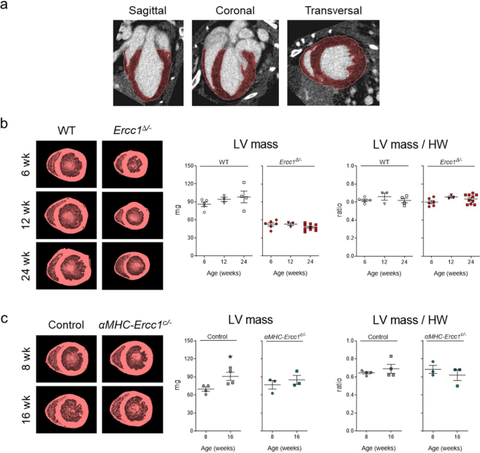
Hybrid Molecular and Functional Micro-CT Imaging Reveals Increased Myocardial Apoptosis Preceding Cardiac Failure in Progeroid Ercc1 Mice.
Purpose: In this study, we explored the role of apoptosis as a potential biomarker for cardiac failure using functional micro-CT and fluorescence molecular tomography (FMT) imaging techniques in Ercc1 mutant mice. Ercc1 is involved in multiple DNA repair pathways, and its mutations contribute to accelerated aging phenotypes in both humans and mice, due to the accumulation of DNA lesions that impair vital DNA functions. We previously found that systemic mutations and cardiomyocyte-restricted deletion of Ercc1 in mice results in left ventricular (LV) dysfunction at older age.
Procedures and results: Here we report that combined functional micro-CT and FMT imaging allowed us to detect apoptosis in systemic Ercc1 mutant mice prior to the development of overt LV dysfunction, suggesting its potential as an early indicator and contributing factor of cardiac impairment. The detection of apoptosis in vivo was feasible as early as 12 weeks of age, even when global LV function appeared normal, underscoring the potential of apoptosis as an early predictor of LV dysfunction, which subsequently manifested at 24 weeks.
Conclusions: This study highlights the utility of combined functional micro-CT and FMT imaging in assessing cardiac function and detecting apoptosis, providing valuable insights into the potential of apoptosis as an early biomarker for cardiac failure.
期刊介绍:
Molecular Imaging and Biology (MIB) invites original contributions (research articles, review articles, commentaries, etc.) on the utilization of molecular imaging (i.e., nuclear imaging, optical imaging, autoradiography and pathology, MRI, MPI, ultrasound imaging, radiomics/genomics etc.) to investigate questions related to biology and health. The objective of MIB is to provide a forum to the discovery of molecular mechanisms of disease through the use of imaging techniques. We aim to investigate the biological nature of disease in patients and establish new molecular imaging diagnostic and therapy procedures.
Some areas that are covered are:
Preclinical and clinical imaging of macromolecular targets (e.g., genes, receptors, enzymes) involved in significant biological processes.
The design, characterization, and study of new molecular imaging probes and contrast agents for the functional interrogation of macromolecular targets.
Development and evaluation of imaging systems including instrumentation, image reconstruction algorithms, image analysis, and display.
Development of molecular assay approaches leading to quantification of the biological information obtained in molecular imaging.
Study of in vivo animal models of disease for the development of new molecular diagnostics and therapeutics.
Extension of in vitro and in vivo discoveries using disease models, into well designed clinical research investigations.
Clinical molecular imaging involving clinical investigations, clinical trials and medical management or cost-effectiveness studies.


 分享
分享

 求助内容:
求助内容: 应助结果提醒方式:
 扫码关注我们
