{"title":"透明增强技术可对通过内镜黏膜下剥离术获得的食管癌进行三维评估。","authors":"Yuichi Asahina, Munetoshi Hinata, Asami Tanaka, Kaori Oshio, Haruki Ogawa, Makoto Aihara, Hiroshi Onodera, Tetsuo Ushiku","doi":"10.1007/s10388-024-01055-x","DOIUrl":null,"url":null,"abstract":"<p><strong>Background: </strong>Although much progress has been made in diagnosis of carcinomas, no established methods have been confirmed to elucidate their morphological features.</p><p><strong>Methods: </strong>Three-dimensional structure of esophageal carcinomas was assessed using transparency-enhancing technology. Endoscopically resected esophageal squamous cell carcinoma was fluorescently stained, optically cleared using a transparency-enhancing reagent called LUCID, and visualized using laser scanning microscopy. The resulting microscope images were converted to virtual HE images for observation using ImageJ software.</p><p><strong>Results: </strong>Microscopic observation and image editing enabled three-dimensional image reconstruction and conversion to virtual HE images. The structure of abnormal blood vessels in esophageal carcinoma recognized by endoscopy could be observed in the 3 dimensions. Squamous cell carcinoma and normal squamous epithelium could be distinguished in the virtual HE images.</p><p><strong>Conclusions: </strong>The results suggested that transparency-enhancing technology and virtual HE images may be feasible for clinical application and represent a novel histopathological method for evaluating endoscopically resected specimens.</p>","PeriodicalId":11918,"journal":{"name":"Esophagus","volume":" ","pages":"405-409"},"PeriodicalIF":3.8000,"publicationDate":"2024-07-01","publicationTypes":"Journal Article","fieldsOfStudy":null,"isOpenAccess":false,"openAccessPdf":"https://www.ncbi.nlm.nih.gov/pmc/articles/PMC11199231/pdf/","citationCount":"0","resultStr":"{\"title\":\"Transparency-enhancing technology allows the three-dimensional assessment of esophageal carcinoma obtained by endoscopic submucosal dissection.\",\"authors\":\"Yuichi Asahina, Munetoshi Hinata, Asami Tanaka, Kaori Oshio, Haruki Ogawa, Makoto Aihara, Hiroshi Onodera, Tetsuo Ushiku\",\"doi\":\"10.1007/s10388-024-01055-x\",\"DOIUrl\":null,\"url\":null,\"abstract\":\"<p><strong>Background: </strong>Although much progress has been made in diagnosis of carcinomas, no established methods have been confirmed to elucidate their morphological features.</p><p><strong>Methods: </strong>Three-dimensional structure of esophageal carcinomas was assessed using transparency-enhancing technology. Endoscopically resected esophageal squamous cell carcinoma was fluorescently stained, optically cleared using a transparency-enhancing reagent called LUCID, and visualized using laser scanning microscopy. The resulting microscope images were converted to virtual HE images for observation using ImageJ software.</p><p><strong>Results: </strong>Microscopic observation and image editing enabled three-dimensional image reconstruction and conversion to virtual HE images. The structure of abnormal blood vessels in esophageal carcinoma recognized by endoscopy could be observed in the 3 dimensions. Squamous cell carcinoma and normal squamous epithelium could be distinguished in the virtual HE images.</p><p><strong>Conclusions: </strong>The results suggested that transparency-enhancing technology and virtual HE images may be feasible for clinical application and represent a novel histopathological method for evaluating endoscopically resected specimens.</p>\",\"PeriodicalId\":11918,\"journal\":{\"name\":\"Esophagus\",\"volume\":\" \",\"pages\":\"405-409\"},\"PeriodicalIF\":3.8000,\"publicationDate\":\"2024-07-01\",\"publicationTypes\":\"Journal Article\",\"fieldsOfStudy\":null,\"isOpenAccess\":false,\"openAccessPdf\":\"https://www.ncbi.nlm.nih.gov/pmc/articles/PMC11199231/pdf/\",\"citationCount\":\"0\",\"resultStr\":null,\"platform\":\"Semanticscholar\",\"paperid\":null,\"PeriodicalName\":\"Esophagus\",\"FirstCategoryId\":\"3\",\"ListUrlMain\":\"https://doi.org/10.1007/s10388-024-01055-x\",\"RegionNum\":4,\"RegionCategory\":\"医学\",\"ArticlePicture\":[],\"TitleCN\":null,\"AbstractTextCN\":null,\"PMCID\":null,\"EPubDate\":\"2024/3/18 0:00:00\",\"PubModel\":\"Epub\",\"JCR\":\"Q3\",\"JCRName\":\"GASTROENTEROLOGY & HEPATOLOGY\",\"Score\":null,\"Total\":0}","platform":"Semanticscholar","paperid":null,"PeriodicalName":"Esophagus","FirstCategoryId":"3","ListUrlMain":"https://doi.org/10.1007/s10388-024-01055-x","RegionNum":4,"RegionCategory":"医学","ArticlePicture":[],"TitleCN":null,"AbstractTextCN":null,"PMCID":null,"EPubDate":"2024/3/18 0:00:00","PubModel":"Epub","JCR":"Q3","JCRName":"GASTROENTEROLOGY & HEPATOLOGY","Score":null,"Total":0}
引用次数: 0
摘要
背景:尽管在诊断食管癌方面取得了很大进展,但还没有确定的方法来阐明食管癌的形态特征:方法:使用透明增强技术评估食管癌的三维结构。对内镜下切除的食管鳞状细胞癌进行荧光染色,使用一种名为 LUCID 的透明增强试剂进行光学清除,然后使用激光扫描显微镜进行观察。使用 ImageJ 软件将显微镜图像转换为虚拟 HE 图像进行观察:结果:显微镜观察和图像编辑实现了三维图像重建并转换为虚拟 HE 图像。结果:通过显微镜观察和图像编辑,可以重建三维图像并转换为虚拟 HE 图像。在虚拟 HE 图像中可以区分鳞状细胞癌和正常鳞状上皮:结果表明,透明增强技术和虚拟 HE 图像在临床应用中是可行的,是评估内镜切除标本的一种新型组织病理学方法。
Transparency-enhancing technology allows the three-dimensional assessment of esophageal carcinoma obtained by endoscopic submucosal dissection.
Background: Although much progress has been made in diagnosis of carcinomas, no established methods have been confirmed to elucidate their morphological features.
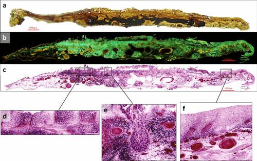
Methods: Three-dimensional structure of esophageal carcinomas was assessed using transparency-enhancing technology. Endoscopically resected esophageal squamous cell carcinoma was fluorescently stained, optically cleared using a transparency-enhancing reagent called LUCID, and visualized using laser scanning microscopy. The resulting microscope images were converted to virtual HE images for observation using ImageJ software.
Results: Microscopic observation and image editing enabled three-dimensional image reconstruction and conversion to virtual HE images. The structure of abnormal blood vessels in esophageal carcinoma recognized by endoscopy could be observed in the 3 dimensions. Squamous cell carcinoma and normal squamous epithelium could be distinguished in the virtual HE images.
Conclusions: The results suggested that transparency-enhancing technology and virtual HE images may be feasible for clinical application and represent a novel histopathological method for evaluating endoscopically resected specimens.
期刊介绍:
Esophagus, the official journal of the Japan Esophageal Society, introduces practitioners and researchers to significant studies in the fields of benign and malignant diseases of the esophagus. The journal welcomes original articles, review articles, and short articles including technical notes ( How I do it ), which will be peer-reviewed by the editorial board. Letters to the editor are also welcome. Special articles on esophageal diseases will be provided by the editorial board, and proceedings of symposia and workshops will be included in special issues for the Annual Congress of the Society.


 分享
分享
 求助内容:
求助内容: 应助结果提醒方式:
 扫码关注我们
