Anhelina Khadanovich, Michal Benes, Radek Kaiser, David Kachlik
{"title":"桡神经浅支穿过上腕管,在伸拇肌和内收肌之间出现,从而为第二指和第三指的桡侧部分提供营养:病例报告和临床意义","authors":"Anhelina Khadanovich, Michal Benes, Radek Kaiser, David Kachlik","doi":"10.1007/s00276-024-03360-7","DOIUrl":null,"url":null,"abstract":"<p>Awareness of unique path of the superficial branch of the radial nerve and its unusual sensory distribution can help avoid potential diagnostic confusion. We present a unique case encountered during a routine dissection of a Central European male cadaver. An unusual course of the superficial branch of the radial nerve was found in the right forearm, where the superficial branch of the radial nerve originated from the radial nerve distally, within the supinator canal, emerged between the extensor digitorum and abductor pollicis longus muscles and supplied the second and a radial half of the third digit, featuring communications with the lateral antebrachial cutaneous nerve and the dorsal branch of the ulnar nerve. Due to dorsal emerging of the superficial branch of the radial nerve the dorsal aspect of the thumb was innervated by the lateral antebrachial cutaneous nerve. To our best knowledge such variation of the superficial branch of the radial nerve has never been reported before. This variation dramatically changes aetiology and manifestation of possible entrapment syndromes which clinicians should be aware of.</p>","PeriodicalId":49296,"journal":{"name":"Surgical and Radiologic Anatomy","volume":"35 1","pages":""},"PeriodicalIF":1.2000,"publicationDate":"2024-04-18","publicationTypes":"Journal Article","fieldsOfStudy":null,"isOpenAccess":false,"openAccessPdf":"","citationCount":"0","resultStr":"{\"title\":\"Superficial branch of the radial nerve passing through the supinator canal, emerging between the extensor digitorum and abductor pollicis longus muscles and consequently supplying the second finger and radial portion of the third finger: a case report and clinical implications\",\"authors\":\"Anhelina Khadanovich, Michal Benes, Radek Kaiser, David Kachlik\",\"doi\":\"10.1007/s00276-024-03360-7\",\"DOIUrl\":null,\"url\":null,\"abstract\":\"<p>Awareness of unique path of the superficial branch of the radial nerve and its unusual sensory distribution can help avoid potential diagnostic confusion. We present a unique case encountered during a routine dissection of a Central European male cadaver. An unusual course of the superficial branch of the radial nerve was found in the right forearm, where the superficial branch of the radial nerve originated from the radial nerve distally, within the supinator canal, emerged between the extensor digitorum and abductor pollicis longus muscles and supplied the second and a radial half of the third digit, featuring communications with the lateral antebrachial cutaneous nerve and the dorsal branch of the ulnar nerve. Due to dorsal emerging of the superficial branch of the radial nerve the dorsal aspect of the thumb was innervated by the lateral antebrachial cutaneous nerve. To our best knowledge such variation of the superficial branch of the radial nerve has never been reported before. This variation dramatically changes aetiology and manifestation of possible entrapment syndromes which clinicians should be aware of.</p>\",\"PeriodicalId\":49296,\"journal\":{\"name\":\"Surgical and Radiologic Anatomy\",\"volume\":\"35 1\",\"pages\":\"\"},\"PeriodicalIF\":1.2000,\"publicationDate\":\"2024-04-18\",\"publicationTypes\":\"Journal Article\",\"fieldsOfStudy\":null,\"isOpenAccess\":false,\"openAccessPdf\":\"\",\"citationCount\":\"0\",\"resultStr\":null,\"platform\":\"Semanticscholar\",\"paperid\":null,\"PeriodicalName\":\"Surgical and Radiologic Anatomy\",\"FirstCategoryId\":\"3\",\"ListUrlMain\":\"https://doi.org/10.1007/s00276-024-03360-7\",\"RegionNum\":4,\"RegionCategory\":\"医学\",\"ArticlePicture\":[],\"TitleCN\":null,\"AbstractTextCN\":null,\"PMCID\":null,\"EPubDate\":\"\",\"PubModel\":\"\",\"JCR\":\"Q3\",\"JCRName\":\"ANATOMY & MORPHOLOGY\",\"Score\":null,\"Total\":0}","platform":"Semanticscholar","paperid":null,"PeriodicalName":"Surgical and Radiologic Anatomy","FirstCategoryId":"3","ListUrlMain":"https://doi.org/10.1007/s00276-024-03360-7","RegionNum":4,"RegionCategory":"医学","ArticlePicture":[],"TitleCN":null,"AbstractTextCN":null,"PMCID":null,"EPubDate":"","PubModel":"","JCR":"Q3","JCRName":"ANATOMY & MORPHOLOGY","Score":null,"Total":0}
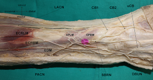
Superficial branch of the radial nerve passing through the supinator canal, emerging between the extensor digitorum and abductor pollicis longus muscles and consequently supplying the second finger and radial portion of the third finger: a case report and clinical implications
Awareness of unique path of the superficial branch of the radial nerve and its unusual sensory distribution can help avoid potential diagnostic confusion. We present a unique case encountered during a routine dissection of a Central European male cadaver. An unusual course of the superficial branch of the radial nerve was found in the right forearm, where the superficial branch of the radial nerve originated from the radial nerve distally, within the supinator canal, emerged between the extensor digitorum and abductor pollicis longus muscles and supplied the second and a radial half of the third digit, featuring communications with the lateral antebrachial cutaneous nerve and the dorsal branch of the ulnar nerve. Due to dorsal emerging of the superficial branch of the radial nerve the dorsal aspect of the thumb was innervated by the lateral antebrachial cutaneous nerve. To our best knowledge such variation of the superficial branch of the radial nerve has never been reported before. This variation dramatically changes aetiology and manifestation of possible entrapment syndromes which clinicians should be aware of.
期刊介绍:
Anatomy is a morphological science which cannot fail to interest the clinician. The practical application of anatomical research to clinical problems necessitates special adaptation and selectivity in choosing from numerous international works. Although there is a tendency to believe that meaningful advances in anatomy are unlikely, constant revision is necessary. Surgical and Radiologic Anatomy, the first international journal of Clinical anatomy has been created in this spirit.
Its goal is to serve clinicians, regardless of speciality-physicians, surgeons, radiologists or other specialists-as an indispensable aid with which they can improve their knowledge of anatomy. Each issue includes: Original papers, review articles, articles on the anatomical bases of medical, surgical and radiological techniques, articles of normal radiologic anatomy, brief reviews of anatomical publications of clinical interest.
Particular attention is given to high quality illustrations, which are indispensable for a better understanding of anatomical problems.
Surgical and Radiologic Anatomy is a journal written by anatomists for clinicians with a special interest in anatomy.


 分享
分享
 求助内容:
求助内容: 应助结果提醒方式:
 扫码关注我们
