Matan Dudaie, Eden Dotan, Itay Barnea, Miki Haifler, Natan T. Shaked
{"title":"利用定量干涉无标记成像流式细胞术检测膀胱癌细胞","authors":"Matan Dudaie, Eden Dotan, Itay Barnea, Miki Haifler, Natan T. Shaked","doi":"10.1002/cyto.a.24846","DOIUrl":null,"url":null,"abstract":"<p>Bladder cancer is one of the most common cancers with a high recurrence rate. Patients undergo mandatory yearly scrutinies, including cystoscopies, which makes bladder cancer highly distressing and costly. Here, we aim to develop a non-invasive, label-free method for the detection of bladder cancer cells in urine samples, which is based on interferometric imaging flow cytometry. Eight urothelial carcinoma and one normal urothelial cell lines, along with red and white blood cells, imaged quantitatively without staining by an interferometric phase microscopy module while flowing in a microfluidic chip, and classified by two machine-learning algorithms, based on deep-learning semantic segmentation convolutional neural network and extreme gradient boosting. Furthermore, urine samples obtained from bladder-cancer patients and healthy volunteers were imaged, and classified by the system. We achieved accuracy and area under the curve (AUC) of 99% and 97% for the cell lines on both machine-learning algorithms. For the real urine samples, the accuracy and AUC were 96% and 96% for the deep-learning algorithm and 95% and 93% for the gradient-boosting algorithm, respectively. By combining label-free interferometric imaging flow cytometry with high-end classification algorithms, we achieved high-performance differentiation between healthy and malignant cells. The proposed technique has the potential to supplant cystoscopy in the bladder cancer surveillance and diagnosis space.</p>","PeriodicalId":11068,"journal":{"name":"Cytometry Part A","volume":"105 8","pages":"570-579"},"PeriodicalIF":2.5000,"publicationDate":"2024-04-26","publicationTypes":"Journal Article","fieldsOfStudy":null,"isOpenAccess":false,"openAccessPdf":"https://onlinelibrary.wiley.com/doi/epdf/10.1002/cyto.a.24846","citationCount":"0","resultStr":"{\"title\":\"Detection of bladder cancer cells using quantitative interferometric label-free imaging flow cytometry\",\"authors\":\"Matan Dudaie, Eden Dotan, Itay Barnea, Miki Haifler, Natan T. Shaked\",\"doi\":\"10.1002/cyto.a.24846\",\"DOIUrl\":null,\"url\":null,\"abstract\":\"<p>Bladder cancer is one of the most common cancers with a high recurrence rate. Patients undergo mandatory yearly scrutinies, including cystoscopies, which makes bladder cancer highly distressing and costly. Here, we aim to develop a non-invasive, label-free method for the detection of bladder cancer cells in urine samples, which is based on interferometric imaging flow cytometry. Eight urothelial carcinoma and one normal urothelial cell lines, along with red and white blood cells, imaged quantitatively without staining by an interferometric phase microscopy module while flowing in a microfluidic chip, and classified by two machine-learning algorithms, based on deep-learning semantic segmentation convolutional neural network and extreme gradient boosting. Furthermore, urine samples obtained from bladder-cancer patients and healthy volunteers were imaged, and classified by the system. We achieved accuracy and area under the curve (AUC) of 99% and 97% for the cell lines on both machine-learning algorithms. For the real urine samples, the accuracy and AUC were 96% and 96% for the deep-learning algorithm and 95% and 93% for the gradient-boosting algorithm, respectively. By combining label-free interferometric imaging flow cytometry with high-end classification algorithms, we achieved high-performance differentiation between healthy and malignant cells. The proposed technique has the potential to supplant cystoscopy in the bladder cancer surveillance and diagnosis space.</p>\",\"PeriodicalId\":11068,\"journal\":{\"name\":\"Cytometry Part A\",\"volume\":\"105 8\",\"pages\":\"570-579\"},\"PeriodicalIF\":2.5000,\"publicationDate\":\"2024-04-26\",\"publicationTypes\":\"Journal Article\",\"fieldsOfStudy\":null,\"isOpenAccess\":false,\"openAccessPdf\":\"https://onlinelibrary.wiley.com/doi/epdf/10.1002/cyto.a.24846\",\"citationCount\":\"0\",\"resultStr\":null,\"platform\":\"Semanticscholar\",\"paperid\":null,\"PeriodicalName\":\"Cytometry Part A\",\"FirstCategoryId\":\"99\",\"ListUrlMain\":\"https://onlinelibrary.wiley.com/doi/10.1002/cyto.a.24846\",\"RegionNum\":4,\"RegionCategory\":\"生物学\",\"ArticlePicture\":[],\"TitleCN\":null,\"AbstractTextCN\":null,\"PMCID\":null,\"EPubDate\":\"\",\"PubModel\":\"\",\"JCR\":\"Q3\",\"JCRName\":\"BIOCHEMICAL RESEARCH METHODS\",\"Score\":null,\"Total\":0}","platform":"Semanticscholar","paperid":null,"PeriodicalName":"Cytometry Part A","FirstCategoryId":"99","ListUrlMain":"https://onlinelibrary.wiley.com/doi/10.1002/cyto.a.24846","RegionNum":4,"RegionCategory":"生物学","ArticlePicture":[],"TitleCN":null,"AbstractTextCN":null,"PMCID":null,"EPubDate":"","PubModel":"","JCR":"Q3","JCRName":"BIOCHEMICAL RESEARCH METHODS","Score":null,"Total":0}
Detection of bladder cancer cells using quantitative interferometric label-free imaging flow cytometry
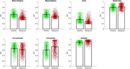
Bladder cancer is one of the most common cancers with a high recurrence rate. Patients undergo mandatory yearly scrutinies, including cystoscopies, which makes bladder cancer highly distressing and costly. Here, we aim to develop a non-invasive, label-free method for the detection of bladder cancer cells in urine samples, which is based on interferometric imaging flow cytometry. Eight urothelial carcinoma and one normal urothelial cell lines, along with red and white blood cells, imaged quantitatively without staining by an interferometric phase microscopy module while flowing in a microfluidic chip, and classified by two machine-learning algorithms, based on deep-learning semantic segmentation convolutional neural network and extreme gradient boosting. Furthermore, urine samples obtained from bladder-cancer patients and healthy volunteers were imaged, and classified by the system. We achieved accuracy and area under the curve (AUC) of 99% and 97% for the cell lines on both machine-learning algorithms. For the real urine samples, the accuracy and AUC were 96% and 96% for the deep-learning algorithm and 95% and 93% for the gradient-boosting algorithm, respectively. By combining label-free interferometric imaging flow cytometry with high-end classification algorithms, we achieved high-performance differentiation between healthy and malignant cells. The proposed technique has the potential to supplant cystoscopy in the bladder cancer surveillance and diagnosis space.
期刊介绍:
Cytometry Part A, the journal of quantitative single-cell analysis, features original research reports and reviews of innovative scientific studies employing quantitative single-cell measurement, separation, manipulation, and modeling techniques, as well as original articles on mechanisms of molecular and cellular functions obtained by cytometry techniques.
The journal welcomes submissions from multiple research fields that fully embrace the study of the cytome:
Biomedical Instrumentation Engineering
Biophotonics
Bioinformatics
Cell Biology
Computational Biology
Data Science
Immunology
Parasitology
Microbiology
Neuroscience
Cancer
Stem Cells
Tissue Regeneration.


 分享
分享
 求助内容:
求助内容: 应助结果提醒方式:
 扫码关注我们
