Takashi Maki, Akira Uchino, Munehiro Sugiyama, Kimiyoshi Mizunuma, Yasutaka Baba
{"title":"通过磁共振血管造影诊断出左侧颈内动脉D型和a型合并缺损","authors":"Takashi Maki, Akira Uchino, Munehiro Sugiyama, Kimiyoshi Mizunuma, Yasutaka Baba","doi":"10.1007/s00276-024-03366-1","DOIUrl":null,"url":null,"abstract":"<h3 data-test=\"abstract-sub-heading\">Purpose</h3><p>To report an unusual case of combined Lie’s types A and D of internal carotid artery (ICA) agenesis, diagnosed by magnetic resonance angiography (MRA).</p><h3 data-test=\"abstract-sub-heading\">Methods</h3><p>A 60-year-old woman with dizziness underwent cranial magnetic resonance imaging (MRI) and MRA of the intracranial region for the evaluation of brain and vascular lesions. The magnetic resonance machine was a 3.0-T scanner.</p><h3 data-test=\"abstract-sub-heading\">Results</h3><p>MRI showed no abnormalities, except for multiple small white matter lesions. MRA showed that the left ICA was absent, except for the supraclinoid segment, and an anastomotic vessel was present between the paraclinoid segments of the bilateral ICAs, indicating Lie’s type D ICA agenesis. The left posterior communicating artery (PCoA) was also present. Thus, there were also features of type A ICA agenesis. The anastomotic vessels between the bilateral ICAs and ipsilateral PCoA were relatively small in caliber.</p><h3 data-test=\"abstract-sub-heading\">Conclusion</h3><p>Lie’s type D ICA agenesis usually does not communicate with the anterior and posterior circulations. We encountered a case of combined type D and type A ICA agenesis. To our knowledge, no similar case has been reported in the English literature. This is the second case of type D ICA agenesis with patent ipsilateral PCoA. We speculate that in case of type A ICA agenesis, when the development of the PCoA is insufficient to support collateral blood flow, an anastomotic vessel between bilateral ICAs may develop.</p>","PeriodicalId":49296,"journal":{"name":"Surgical and Radiologic Anatomy","volume":"45 1","pages":""},"PeriodicalIF":1.2000,"publicationDate":"2024-04-29","publicationTypes":"Journal Article","fieldsOfStudy":null,"isOpenAccess":false,"openAccessPdf":"","citationCount":"0","resultStr":"{\"title\":\"Combined lie’s type D and type a agenesis of the left internal carotid artery diagnosed by magnetic resonance angiography\",\"authors\":\"Takashi Maki, Akira Uchino, Munehiro Sugiyama, Kimiyoshi Mizunuma, Yasutaka Baba\",\"doi\":\"10.1007/s00276-024-03366-1\",\"DOIUrl\":null,\"url\":null,\"abstract\":\"<h3 data-test=\\\"abstract-sub-heading\\\">Purpose</h3><p>To report an unusual case of combined Lie’s types A and D of internal carotid artery (ICA) agenesis, diagnosed by magnetic resonance angiography (MRA).</p><h3 data-test=\\\"abstract-sub-heading\\\">Methods</h3><p>A 60-year-old woman with dizziness underwent cranial magnetic resonance imaging (MRI) and MRA of the intracranial region for the evaluation of brain and vascular lesions. The magnetic resonance machine was a 3.0-T scanner.</p><h3 data-test=\\\"abstract-sub-heading\\\">Results</h3><p>MRI showed no abnormalities, except for multiple small white matter lesions. MRA showed that the left ICA was absent, except for the supraclinoid segment, and an anastomotic vessel was present between the paraclinoid segments of the bilateral ICAs, indicating Lie’s type D ICA agenesis. The left posterior communicating artery (PCoA) was also present. Thus, there were also features of type A ICA agenesis. The anastomotic vessels between the bilateral ICAs and ipsilateral PCoA were relatively small in caliber.</p><h3 data-test=\\\"abstract-sub-heading\\\">Conclusion</h3><p>Lie’s type D ICA agenesis usually does not communicate with the anterior and posterior circulations. We encountered a case of combined type D and type A ICA agenesis. To our knowledge, no similar case has been reported in the English literature. This is the second case of type D ICA agenesis with patent ipsilateral PCoA. We speculate that in case of type A ICA agenesis, when the development of the PCoA is insufficient to support collateral blood flow, an anastomotic vessel between bilateral ICAs may develop.</p>\",\"PeriodicalId\":49296,\"journal\":{\"name\":\"Surgical and Radiologic Anatomy\",\"volume\":\"45 1\",\"pages\":\"\"},\"PeriodicalIF\":1.2000,\"publicationDate\":\"2024-04-29\",\"publicationTypes\":\"Journal Article\",\"fieldsOfStudy\":null,\"isOpenAccess\":false,\"openAccessPdf\":\"\",\"citationCount\":\"0\",\"resultStr\":null,\"platform\":\"Semanticscholar\",\"paperid\":null,\"PeriodicalName\":\"Surgical and Radiologic Anatomy\",\"FirstCategoryId\":\"3\",\"ListUrlMain\":\"https://doi.org/10.1007/s00276-024-03366-1\",\"RegionNum\":4,\"RegionCategory\":\"医学\",\"ArticlePicture\":[],\"TitleCN\":null,\"AbstractTextCN\":null,\"PMCID\":null,\"EPubDate\":\"\",\"PubModel\":\"\",\"JCR\":\"Q3\",\"JCRName\":\"ANATOMY & MORPHOLOGY\",\"Score\":null,\"Total\":0}","platform":"Semanticscholar","paperid":null,"PeriodicalName":"Surgical and Radiologic Anatomy","FirstCategoryId":"3","ListUrlMain":"https://doi.org/10.1007/s00276-024-03366-1","RegionNum":4,"RegionCategory":"医学","ArticlePicture":[],"TitleCN":null,"AbstractTextCN":null,"PMCID":null,"EPubDate":"","PubModel":"","JCR":"Q3","JCRName":"ANATOMY & MORPHOLOGY","Score":null,"Total":0}
引用次数: 0
摘要
目的 通过磁共振血管造影术(MRA)确诊一例罕见的 A 型和 D 型合并颈内动脉(ICA)缺失病例。方法 一位头晕的 60 岁女性接受了头颅磁共振成像(MRI)和颅内磁共振血管造影术,以评估大脑和血管病变。磁共振成像仪是一台 3.0 T 扫描仪。MRA显示,左侧ICA除上盲段外缺失,双侧ICA的旁盲段之间存在吻合血管,表明Lie's D型ICA缺失。左后交通动脉(PCoA)也存在。因此,也有 A 型 ICA 成长的特征。双侧 ICA 与同侧 PCoA 之间的吻合血管口径相对较小。我们遇到了一例合并 D 型和 A 型室内动脉缺失的病例。据我们所知,英文文献中还没有类似病例的报道。这是第二例同侧 PCoA 成通畅的 D 型 ICA 成长病例。我们推测,在 A 型内动脉缺失的病例中,当 PCoA 的发育不足以支持侧支血流时,双侧内动脉之间可能会出现吻合血管。
Combined lie’s type D and type a agenesis of the left internal carotid artery diagnosed by magnetic resonance angiography
Purpose
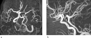
To report an unusual case of combined Lie’s types A and D of internal carotid artery (ICA) agenesis, diagnosed by magnetic resonance angiography (MRA).
Methods
A 60-year-old woman with dizziness underwent cranial magnetic resonance imaging (MRI) and MRA of the intracranial region for the evaluation of brain and vascular lesions. The magnetic resonance machine was a 3.0-T scanner.
Results
MRI showed no abnormalities, except for multiple small white matter lesions. MRA showed that the left ICA was absent, except for the supraclinoid segment, and an anastomotic vessel was present between the paraclinoid segments of the bilateral ICAs, indicating Lie’s type D ICA agenesis. The left posterior communicating artery (PCoA) was also present. Thus, there were also features of type A ICA agenesis. The anastomotic vessels between the bilateral ICAs and ipsilateral PCoA were relatively small in caliber.
Conclusion
Lie’s type D ICA agenesis usually does not communicate with the anterior and posterior circulations. We encountered a case of combined type D and type A ICA agenesis. To our knowledge, no similar case has been reported in the English literature. This is the second case of type D ICA agenesis with patent ipsilateral PCoA. We speculate that in case of type A ICA agenesis, when the development of the PCoA is insufficient to support collateral blood flow, an anastomotic vessel between bilateral ICAs may develop.
期刊介绍:
Anatomy is a morphological science which cannot fail to interest the clinician. The practical application of anatomical research to clinical problems necessitates special adaptation and selectivity in choosing from numerous international works. Although there is a tendency to believe that meaningful advances in anatomy are unlikely, constant revision is necessary. Surgical and Radiologic Anatomy, the first international journal of Clinical anatomy has been created in this spirit.
Its goal is to serve clinicians, regardless of speciality-physicians, surgeons, radiologists or other specialists-as an indispensable aid with which they can improve their knowledge of anatomy. Each issue includes: Original papers, review articles, articles on the anatomical bases of medical, surgical and radiological techniques, articles of normal radiologic anatomy, brief reviews of anatomical publications of clinical interest.
Particular attention is given to high quality illustrations, which are indispensable for a better understanding of anatomical problems.
Surgical and Radiologic Anatomy is a journal written by anatomists for clinicians with a special interest in anatomy.


 分享
分享
 求助内容:
求助内容: 应助结果提醒方式:
 扫码关注我们
