Aria M Salyapongse, Jeffrey P Kanne, Prashant Nagpal, Nicholas C Laucis, B Keegan Markhardt, Zhye Yin, Scott Slavic, Meghan G Lubner, Timothy P Szczykutowicz
{"title":"能量积分和深硅光子计数 CT 的空间分辨率保真度比较:对肺部成像的影响","authors":"Aria M Salyapongse, Jeffrey P Kanne, Prashant Nagpal, Nicholas C Laucis, B Keegan Markhardt, Zhye Yin, Scott Slavic, Meghan G Lubner, Timothy P Szczykutowicz","doi":"10.1097/RTI.0000000000000788","DOIUrl":null,"url":null,"abstract":"<p><strong>Purpose: </strong>We investigated spatial resolution loss away from isocenter for a prototype deep silicon photon-counting detector (PCD) CT scanner and compare with a clinical energy-integrating detector (EID) CT scanner.</p><p><strong>Materials and methods: </strong>We performed three scans on a wire phantom at four positions (isocenter, 6.7, 11.8, and 17.1 cm off isocenter). The acquisition modes were 120 kV EID CT, 120 kV high-definition (HD) EID CT, and 120 kV PCD CT. HD mode used double the projection view angles per rotation as the \"regular\" EID scan mode. The diameter of the wire was calculated by taking the full width of half max (FWHM) of a profile drawn over the radial and azimuthal directions of the wire. Change in wire diameter appearance was assessed by calculating the ratio of the radial and azimuthal diameter relative to isocenter. t tests were used to make pairwise comparisons of the wire diameter ratio with each acquisition and mean ratios' difference from unity.</p><p><strong>Results: </strong>Deep silicon PCD CT had statistically smaller ( P <0.05) changes in diameter ratio for both radial and azimuthal directions compared with both regular and HD EID modes and was not statistically different from unity ( P <0.05). Maximum increases in FWMH relative to isocenter were 36%, 12%, and 1% for regular EID, HD EID, and deep silicon PCD, respectively.</p><p><strong>Conclusion: </strong>Deep silicon PCD CT exhibits less change in spatial resolution in both the radial and azimuthal directions compared with EID CT.</p>","PeriodicalId":49974,"journal":{"name":"Journal of Thoracic Imaging","volume":" ","pages":"344-350"},"PeriodicalIF":1.9000,"publicationDate":"2024-11-01","publicationTypes":"Journal Article","fieldsOfStudy":null,"isOpenAccess":false,"openAccessPdf":"https://www.ncbi.nlm.nih.gov/pmc/articles/PMC11495528/pdf/","citationCount":"0","resultStr":"{\"title\":\"Spatial Resolution Fidelity Comparison Between Energy Integrating and Deep Silicon Photon Counting CT: Implications for Pulmonary Imaging.\",\"authors\":\"Aria M Salyapongse, Jeffrey P Kanne, Prashant Nagpal, Nicholas C Laucis, B Keegan Markhardt, Zhye Yin, Scott Slavic, Meghan G Lubner, Timothy P Szczykutowicz\",\"doi\":\"10.1097/RTI.0000000000000788\",\"DOIUrl\":null,\"url\":null,\"abstract\":\"<p><strong>Purpose: </strong>We investigated spatial resolution loss away from isocenter for a prototype deep silicon photon-counting detector (PCD) CT scanner and compare with a clinical energy-integrating detector (EID) CT scanner.</p><p><strong>Materials and methods: </strong>We performed three scans on a wire phantom at four positions (isocenter, 6.7, 11.8, and 17.1 cm off isocenter). The acquisition modes were 120 kV EID CT, 120 kV high-definition (HD) EID CT, and 120 kV PCD CT. HD mode used double the projection view angles per rotation as the \\\"regular\\\" EID scan mode. The diameter of the wire was calculated by taking the full width of half max (FWHM) of a profile drawn over the radial and azimuthal directions of the wire. Change in wire diameter appearance was assessed by calculating the ratio of the radial and azimuthal diameter relative to isocenter. t tests were used to make pairwise comparisons of the wire diameter ratio with each acquisition and mean ratios' difference from unity.</p><p><strong>Results: </strong>Deep silicon PCD CT had statistically smaller ( P <0.05) changes in diameter ratio for both radial and azimuthal directions compared with both regular and HD EID modes and was not statistically different from unity ( P <0.05). Maximum increases in FWMH relative to isocenter were 36%, 12%, and 1% for regular EID, HD EID, and deep silicon PCD, respectively.</p><p><strong>Conclusion: </strong>Deep silicon PCD CT exhibits less change in spatial resolution in both the radial and azimuthal directions compared with EID CT.</p>\",\"PeriodicalId\":49974,\"journal\":{\"name\":\"Journal of Thoracic Imaging\",\"volume\":\" \",\"pages\":\"344-350\"},\"PeriodicalIF\":1.9000,\"publicationDate\":\"2024-11-01\",\"publicationTypes\":\"Journal Article\",\"fieldsOfStudy\":null,\"isOpenAccess\":false,\"openAccessPdf\":\"https://www.ncbi.nlm.nih.gov/pmc/articles/PMC11495528/pdf/\",\"citationCount\":\"0\",\"resultStr\":null,\"platform\":\"Semanticscholar\",\"paperid\":null,\"PeriodicalName\":\"Journal of Thoracic Imaging\",\"FirstCategoryId\":\"3\",\"ListUrlMain\":\"https://doi.org/10.1097/RTI.0000000000000788\",\"RegionNum\":4,\"RegionCategory\":\"医学\",\"ArticlePicture\":[],\"TitleCN\":null,\"AbstractTextCN\":null,\"PMCID\":null,\"EPubDate\":\"2024/5/7 0:00:00\",\"PubModel\":\"Epub\",\"JCR\":\"Q3\",\"JCRName\":\"RADIOLOGY, NUCLEAR MEDICINE & MEDICAL IMAGING\",\"Score\":null,\"Total\":0}","platform":"Semanticscholar","paperid":null,"PeriodicalName":"Journal of Thoracic Imaging","FirstCategoryId":"3","ListUrlMain":"https://doi.org/10.1097/RTI.0000000000000788","RegionNum":4,"RegionCategory":"医学","ArticlePicture":[],"TitleCN":null,"AbstractTextCN":null,"PMCID":null,"EPubDate":"2024/5/7 0:00:00","PubModel":"Epub","JCR":"Q3","JCRName":"RADIOLOGY, NUCLEAR MEDICINE & MEDICAL IMAGING","Score":null,"Total":0}
Spatial Resolution Fidelity Comparison Between Energy Integrating and Deep Silicon Photon Counting CT: Implications for Pulmonary Imaging.
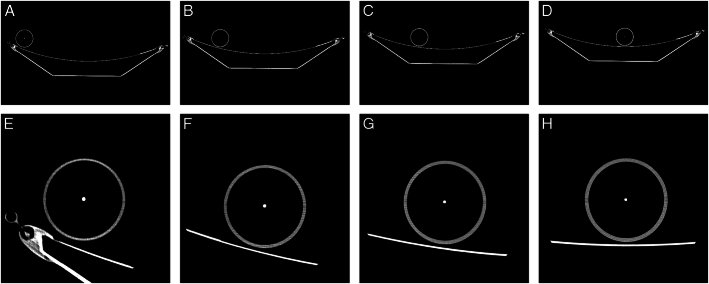
Purpose: We investigated spatial resolution loss away from isocenter for a prototype deep silicon photon-counting detector (PCD) CT scanner and compare with a clinical energy-integrating detector (EID) CT scanner.
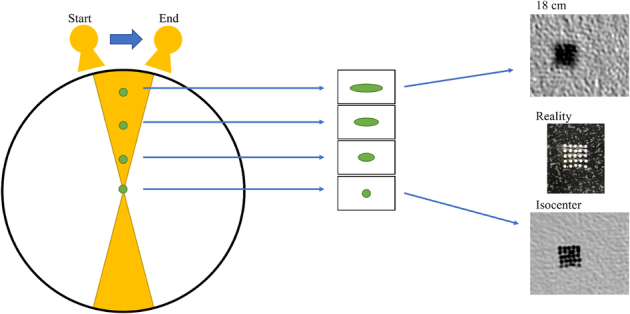
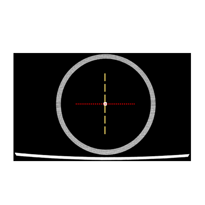
Materials and methods: We performed three scans on a wire phantom at four positions (isocenter, 6.7, 11.8, and 17.1 cm off isocenter). The acquisition modes were 120 kV EID CT, 120 kV high-definition (HD) EID CT, and 120 kV PCD CT. HD mode used double the projection view angles per rotation as the "regular" EID scan mode. The diameter of the wire was calculated by taking the full width of half max (FWHM) of a profile drawn over the radial and azimuthal directions of the wire. Change in wire diameter appearance was assessed by calculating the ratio of the radial and azimuthal diameter relative to isocenter. t tests were used to make pairwise comparisons of the wire diameter ratio with each acquisition and mean ratios' difference from unity.
Results: Deep silicon PCD CT had statistically smaller ( P <0.05) changes in diameter ratio for both radial and azimuthal directions compared with both regular and HD EID modes and was not statistically different from unity ( P <0.05). Maximum increases in FWMH relative to isocenter were 36%, 12%, and 1% for regular EID, HD EID, and deep silicon PCD, respectively.
Conclusion: Deep silicon PCD CT exhibits less change in spatial resolution in both the radial and azimuthal directions compared with EID CT.
期刊介绍:
Journal of Thoracic Imaging (JTI) provides authoritative information on all aspects of the use of imaging techniques in the diagnosis of cardiac and pulmonary diseases. Original articles and analytical reviews published in this timely journal provide the very latest thinking of leading experts concerning the use of chest radiography, computed tomography, magnetic resonance imaging, positron emission tomography, ultrasound, and all other promising imaging techniques in cardiopulmonary radiology.
Official Journal of the Society of Thoracic Radiology:
Japanese Society of Thoracic Radiology
Korean Society of Thoracic Radiology
European Society of Thoracic Imaging.


 分享
分享
 求助内容:
求助内容: 应助结果提醒方式:
 扫码关注我们
