{"title":"坏死牙髓和根尖牙周炎恒牙的血管再造术","authors":"Wala Saad, Manal Almaslamani, Abdul Rahman Saleh","doi":"10.2147/CCIDE.S464214","DOIUrl":null,"url":null,"abstract":"<p><p>This case report details a pulp revascularization treatment administered to a mature permanent tooth exhibiting pulp necrosis. A 22-years old female patient complained of the recurrence of a sinus tract labial of the maxillary right central incisor; which was tender on biting. Diagnosis of pulp necrosis and symptomatic apical periodontitis. Preoperative periapical and CBCT radiographs showed root with wide apical foramen and large apical radiolucency. Pulp revascularization procedure was performed using 1.3% sodium hypochlorite irrigation, 17% Ethylenediaminetetraacetic acid irrigation, and calcium hydroxide intracanal dressing for 2 weeks. During the last visit, intentional bleeding was induced, collagen matrix was set over the blood clot, 2 mm of mineral trioxide aggregate and glass-ionomer filling was placed. A year of follow-up, the tooth showed no signs or symptoms and responded normally to the sensibility tests. Intra-oral periapical radiograph and the CBCT showed significant reduction in the periapical lesion's size, slight reduction in the apical foramen's size, and hard radiopaque material deposition at the root's middle third.</p>","PeriodicalId":10445,"journal":{"name":"Clinical, Cosmetic and Investigational Dentistry","volume":"16 ","pages":"227-235"},"PeriodicalIF":1.8000,"publicationDate":"2024-06-18","publicationTypes":"Journal Article","fieldsOfStudy":null,"isOpenAccess":false,"openAccessPdf":"https://www.ncbi.nlm.nih.gov/pmc/articles/PMC11193457/pdf/","citationCount":"0","resultStr":"{\"title\":\"Revascularization of a Permanent Tooth with Necrotic Pulp and Apical Periodontitis.\",\"authors\":\"Wala Saad, Manal Almaslamani, Abdul Rahman Saleh\",\"doi\":\"10.2147/CCIDE.S464214\",\"DOIUrl\":null,\"url\":null,\"abstract\":\"<p><p>This case report details a pulp revascularization treatment administered to a mature permanent tooth exhibiting pulp necrosis. A 22-years old female patient complained of the recurrence of a sinus tract labial of the maxillary right central incisor; which was tender on biting. Diagnosis of pulp necrosis and symptomatic apical periodontitis. Preoperative periapical and CBCT radiographs showed root with wide apical foramen and large apical radiolucency. Pulp revascularization procedure was performed using 1.3% sodium hypochlorite irrigation, 17% Ethylenediaminetetraacetic acid irrigation, and calcium hydroxide intracanal dressing for 2 weeks. During the last visit, intentional bleeding was induced, collagen matrix was set over the blood clot, 2 mm of mineral trioxide aggregate and glass-ionomer filling was placed. A year of follow-up, the tooth showed no signs or symptoms and responded normally to the sensibility tests. Intra-oral periapical radiograph and the CBCT showed significant reduction in the periapical lesion's size, slight reduction in the apical foramen's size, and hard radiopaque material deposition at the root's middle third.</p>\",\"PeriodicalId\":10445,\"journal\":{\"name\":\"Clinical, Cosmetic and Investigational Dentistry\",\"volume\":\"16 \",\"pages\":\"227-235\"},\"PeriodicalIF\":1.8000,\"publicationDate\":\"2024-06-18\",\"publicationTypes\":\"Journal Article\",\"fieldsOfStudy\":null,\"isOpenAccess\":false,\"openAccessPdf\":\"https://www.ncbi.nlm.nih.gov/pmc/articles/PMC11193457/pdf/\",\"citationCount\":\"0\",\"resultStr\":null,\"platform\":\"Semanticscholar\",\"paperid\":null,\"PeriodicalName\":\"Clinical, Cosmetic and Investigational Dentistry\",\"FirstCategoryId\":\"1085\",\"ListUrlMain\":\"https://doi.org/10.2147/CCIDE.S464214\",\"RegionNum\":0,\"RegionCategory\":null,\"ArticlePicture\":[],\"TitleCN\":null,\"AbstractTextCN\":null,\"PMCID\":null,\"EPubDate\":\"2024/1/1 0:00:00\",\"PubModel\":\"eCollection\",\"JCR\":\"Q3\",\"JCRName\":\"DENTISTRY, ORAL SURGERY & MEDICINE\",\"Score\":null,\"Total\":0}","platform":"Semanticscholar","paperid":null,"PeriodicalName":"Clinical, Cosmetic and Investigational Dentistry","FirstCategoryId":"1085","ListUrlMain":"https://doi.org/10.2147/CCIDE.S464214","RegionNum":0,"RegionCategory":null,"ArticlePicture":[],"TitleCN":null,"AbstractTextCN":null,"PMCID":null,"EPubDate":"2024/1/1 0:00:00","PubModel":"eCollection","JCR":"Q3","JCRName":"DENTISTRY, ORAL SURGERY & MEDICINE","Score":null,"Total":0}
引用次数: 0
摘要
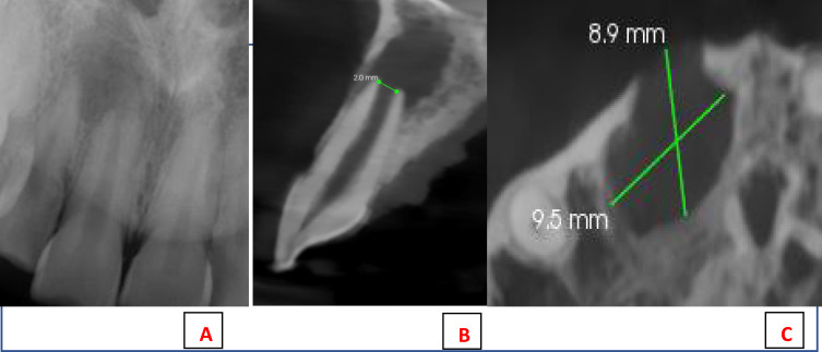
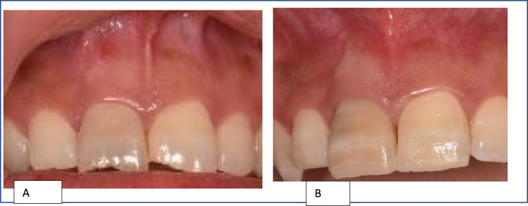
本病例报告详细介绍了对一颗牙髓坏死的成熟恒牙进行的牙髓再造治疗。一名 22 岁的女性患者主诉上颌右中切牙唇侧窦道复发,咬合时有触痛。诊断为牙髓坏死和无症状根尖牙周炎。术前根尖周炎和 CBCT X 光片显示,牙根根尖孔宽大,根尖有较大的放射斑。使用1.3%次氯酸钠冲洗、17%乙二胺四乙酸冲洗和氢氧化钙敷料进行了为期两周的牙髓再通术。在最后一次就诊时,诱导了故意出血,在血凝块上设置了胶原基质,放置了 2 毫米的三氧化二矿骨料和玻璃-离子填充物。随访一年后,牙齿没有出现任何体征或症状,对感度测试的反应也很正常。口内根尖周炎X光片和CBCT显示,根尖周炎病灶明显缩小,根尖孔略有缩小,牙根中间三分之一处有不透射线的硬质材料沉积。
Revascularization of a Permanent Tooth with Necrotic Pulp and Apical Periodontitis.
This case report details a pulp revascularization treatment administered to a mature permanent tooth exhibiting pulp necrosis. A 22-years old female patient complained of the recurrence of a sinus tract labial of the maxillary right central incisor; which was tender on biting. Diagnosis of pulp necrosis and symptomatic apical periodontitis. Preoperative periapical and CBCT radiographs showed root with wide apical foramen and large apical radiolucency. Pulp revascularization procedure was performed using 1.3% sodium hypochlorite irrigation, 17% Ethylenediaminetetraacetic acid irrigation, and calcium hydroxide intracanal dressing for 2 weeks. During the last visit, intentional bleeding was induced, collagen matrix was set over the blood clot, 2 mm of mineral trioxide aggregate and glass-ionomer filling was placed. A year of follow-up, the tooth showed no signs or symptoms and responded normally to the sensibility tests. Intra-oral periapical radiograph and the CBCT showed significant reduction in the periapical lesion's size, slight reduction in the apical foramen's size, and hard radiopaque material deposition at the root's middle third.


 分享
分享
 求助内容:
求助内容: 应助结果提醒方式:
 扫码关注我们
