{"title":"将 S-(3-[18F]氟丙基)-D-高半胱氨酸和 O-(2-[18F]氟乙基)-D-酪氨酸作为细菌特异性放射性同位素用于 PET 感染成像的试点评估。","authors":"Helen M Betts, Jeni C Luckett, Philip J Hill","doi":"10.1007/s11307-024-01929-7","DOIUrl":null,"url":null,"abstract":"<p><strong>Purpose: </strong>There is currently no ideal radiotracer for imaging bacterial infections. Radiolabelled D-amino acids are promising candidates because they are actively incorporated into the peptidoglycan of the bacterial cell wall, a structural feature which is absent in human cells. This work describes fluorine-18 labelled analogues of D-tyrosine and D-methionine, O-(2-[<sup>18</sup>F]fluoroethyl)-D-tyrosine (D-[<sup>18</sup>F]FET) and S-(3-[<sup>18</sup>F]fluoropropyl)-D-homocysteine (D-[<sup>18</sup>F]FPHCys), and their pilot evaluation studies as potential radiotracers for imaging bacterial infection.</p><p><strong>Procedures: </strong>D-[<sup>18</sup>F]FET and D-[<sup>18</sup>F]FPHCys were prepared in classical fluorination-deprotection reactions, and their uptake in Staphylococcus aureus and Pseudomonas aeruginosa was evaluated over 2 h. Heat killed bacteria were used as controls. A clinically-relevant foreign body model of S. aureus infection was established in Balb/c mice, as well as a sterile foreign body to mimic inflammation. The ex vivo biodistribution of D-[<sup>18</sup>F]FPHCys in the infected and inflamed mice was evaluated after 1 h, by dissection and gamma counting. The uptake was compared to that of [<sup>18</sup>F]FDG.</p><p><strong>Results: </strong>In vitro uptake of both D-[<sup>18</sup>F]FET and D-[<sup>18</sup>F]FPHCys was specific to live bacteria. Uptake was higher in S. aureus than in P. aeruginosa for both radiotracers, and of the two, higher for D-[<sup>18</sup>F]FPHCys than D-[<sup>18</sup>F]FET. Blocking experiments with non-radioactive D-[<sup>19</sup>F]FPHCys confirmed specificity of uptake. In vivo, D-[<sup>18</sup>F]FPHCys had greater accumulation in S. aureus infection compared with sterile inflammation, which was statistically significant. As anticipated, [<sup>18</sup>F]FDG showed no significant difference in uptake between infection and inflammation.</p><p><strong>Conclusions: </strong>D-[<sup>18</sup>F]FPHCys uptake was higher in infected tissues than inflammation, and represents a fluorine-18 labelled D-AA with potential to detect a S. aureus reference strain (Xen29) in vivo. Additional studies are needed to evaluate uptake of this radiotracer in clinical isolates.</p>","PeriodicalId":18760,"journal":{"name":"Molecular Imaging and Biology","volume":" ","pages":"704-713"},"PeriodicalIF":2.5000,"publicationDate":"2024-08-01","publicationTypes":"Journal Article","fieldsOfStudy":null,"isOpenAccess":false,"openAccessPdf":"https://www.ncbi.nlm.nih.gov/pmc/articles/PMC11282134/pdf/","citationCount":"0","resultStr":"{\"title\":\"Pilot Evaluation of S-(3-[<sup>18</sup>F]Fluoropropyl)-D-Homocysteine and O-(2-[<sup>18</sup>F]Fluoroethyl)-D-Tyrosine as Bacteria-Specific Radiotracers for PET Imaging of Infection.\",\"authors\":\"Helen M Betts, Jeni C Luckett, Philip J Hill\",\"doi\":\"10.1007/s11307-024-01929-7\",\"DOIUrl\":null,\"url\":null,\"abstract\":\"<p><strong>Purpose: </strong>There is currently no ideal radiotracer for imaging bacterial infections. Radiolabelled D-amino acids are promising candidates because they are actively incorporated into the peptidoglycan of the bacterial cell wall, a structural feature which is absent in human cells. This work describes fluorine-18 labelled analogues of D-tyrosine and D-methionine, O-(2-[<sup>18</sup>F]fluoroethyl)-D-tyrosine (D-[<sup>18</sup>F]FET) and S-(3-[<sup>18</sup>F]fluoropropyl)-D-homocysteine (D-[<sup>18</sup>F]FPHCys), and their pilot evaluation studies as potential radiotracers for imaging bacterial infection.</p><p><strong>Procedures: </strong>D-[<sup>18</sup>F]FET and D-[<sup>18</sup>F]FPHCys were prepared in classical fluorination-deprotection reactions, and their uptake in Staphylococcus aureus and Pseudomonas aeruginosa was evaluated over 2 h. Heat killed bacteria were used as controls. A clinically-relevant foreign body model of S. aureus infection was established in Balb/c mice, as well as a sterile foreign body to mimic inflammation. The ex vivo biodistribution of D-[<sup>18</sup>F]FPHCys in the infected and inflamed mice was evaluated after 1 h, by dissection and gamma counting. The uptake was compared to that of [<sup>18</sup>F]FDG.</p><p><strong>Results: </strong>In vitro uptake of both D-[<sup>18</sup>F]FET and D-[<sup>18</sup>F]FPHCys was specific to live bacteria. Uptake was higher in S. aureus than in P. aeruginosa for both radiotracers, and of the two, higher for D-[<sup>18</sup>F]FPHCys than D-[<sup>18</sup>F]FET. Blocking experiments with non-radioactive D-[<sup>19</sup>F]FPHCys confirmed specificity of uptake. In vivo, D-[<sup>18</sup>F]FPHCys had greater accumulation in S. aureus infection compared with sterile inflammation, which was statistically significant. As anticipated, [<sup>18</sup>F]FDG showed no significant difference in uptake between infection and inflammation.</p><p><strong>Conclusions: </strong>D-[<sup>18</sup>F]FPHCys uptake was higher in infected tissues than inflammation, and represents a fluorine-18 labelled D-AA with potential to detect a S. aureus reference strain (Xen29) in vivo. Additional studies are needed to evaluate uptake of this radiotracer in clinical isolates.</p>\",\"PeriodicalId\":18760,\"journal\":{\"name\":\"Molecular Imaging and Biology\",\"volume\":\" \",\"pages\":\"704-713\"},\"PeriodicalIF\":2.5000,\"publicationDate\":\"2024-08-01\",\"publicationTypes\":\"Journal Article\",\"fieldsOfStudy\":null,\"isOpenAccess\":false,\"openAccessPdf\":\"https://www.ncbi.nlm.nih.gov/pmc/articles/PMC11282134/pdf/\",\"citationCount\":\"0\",\"resultStr\":null,\"platform\":\"Semanticscholar\",\"paperid\":null,\"PeriodicalName\":\"Molecular Imaging and Biology\",\"FirstCategoryId\":\"3\",\"ListUrlMain\":\"https://doi.org/10.1007/s11307-024-01929-7\",\"RegionNum\":4,\"RegionCategory\":\"医学\",\"ArticlePicture\":[],\"TitleCN\":null,\"AbstractTextCN\":null,\"PMCID\":null,\"EPubDate\":\"2024/6/28 0:00:00\",\"PubModel\":\"Epub\",\"JCR\":\"Q2\",\"JCRName\":\"RADIOLOGY, NUCLEAR MEDICINE & MEDICAL IMAGING\",\"Score\":null,\"Total\":0}","platform":"Semanticscholar","paperid":null,"PeriodicalName":"Molecular Imaging and Biology","FirstCategoryId":"3","ListUrlMain":"https://doi.org/10.1007/s11307-024-01929-7","RegionNum":4,"RegionCategory":"医学","ArticlePicture":[],"TitleCN":null,"AbstractTextCN":null,"PMCID":null,"EPubDate":"2024/6/28 0:00:00","PubModel":"Epub","JCR":"Q2","JCRName":"RADIOLOGY, NUCLEAR MEDICINE & MEDICAL IMAGING","Score":null,"Total":0}
Pilot Evaluation of S-(3-[18F]Fluoropropyl)-D-Homocysteine and O-(2-[18F]Fluoroethyl)-D-Tyrosine as Bacteria-Specific Radiotracers for PET Imaging of Infection.
Purpose: There is currently no ideal radiotracer for imaging bacterial infections. Radiolabelled D-amino acids are promising candidates because they are actively incorporated into the peptidoglycan of the bacterial cell wall, a structural feature which is absent in human cells. This work describes fluorine-18 labelled analogues of D-tyrosine and D-methionine, O-(2-[18F]fluoroethyl)-D-tyrosine (D-[18F]FET) and S-(3-[18F]fluoropropyl)-D-homocysteine (D-[18F]FPHCys), and their pilot evaluation studies as potential radiotracers for imaging bacterial infection.
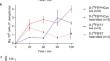
Procedures: D-[18F]FET and D-[18F]FPHCys were prepared in classical fluorination-deprotection reactions, and their uptake in Staphylococcus aureus and Pseudomonas aeruginosa was evaluated over 2 h. Heat killed bacteria were used as controls. A clinically-relevant foreign body model of S. aureus infection was established in Balb/c mice, as well as a sterile foreign body to mimic inflammation. The ex vivo biodistribution of D-[18F]FPHCys in the infected and inflamed mice was evaluated after 1 h, by dissection and gamma counting. The uptake was compared to that of [18F]FDG.
Results: In vitro uptake of both D-[18F]FET and D-[18F]FPHCys was specific to live bacteria. Uptake was higher in S. aureus than in P. aeruginosa for both radiotracers, and of the two, higher for D-[18F]FPHCys than D-[18F]FET. Blocking experiments with non-radioactive D-[19F]FPHCys confirmed specificity of uptake. In vivo, D-[18F]FPHCys had greater accumulation in S. aureus infection compared with sterile inflammation, which was statistically significant. As anticipated, [18F]FDG showed no significant difference in uptake between infection and inflammation.
Conclusions: D-[18F]FPHCys uptake was higher in infected tissues than inflammation, and represents a fluorine-18 labelled D-AA with potential to detect a S. aureus reference strain (Xen29) in vivo. Additional studies are needed to evaluate uptake of this radiotracer in clinical isolates.
期刊介绍:
Molecular Imaging and Biology (MIB) invites original contributions (research articles, review articles, commentaries, etc.) on the utilization of molecular imaging (i.e., nuclear imaging, optical imaging, autoradiography and pathology, MRI, MPI, ultrasound imaging, radiomics/genomics etc.) to investigate questions related to biology and health. The objective of MIB is to provide a forum to the discovery of molecular mechanisms of disease through the use of imaging techniques. We aim to investigate the biological nature of disease in patients and establish new molecular imaging diagnostic and therapy procedures.
Some areas that are covered are:
Preclinical and clinical imaging of macromolecular targets (e.g., genes, receptors, enzymes) involved in significant biological processes.
The design, characterization, and study of new molecular imaging probes and contrast agents for the functional interrogation of macromolecular targets.
Development and evaluation of imaging systems including instrumentation, image reconstruction algorithms, image analysis, and display.
Development of molecular assay approaches leading to quantification of the biological information obtained in molecular imaging.
Study of in vivo animal models of disease for the development of new molecular diagnostics and therapeutics.
Extension of in vitro and in vivo discoveries using disease models, into well designed clinical research investigations.
Clinical molecular imaging involving clinical investigations, clinical trials and medical management or cost-effectiveness studies.


 分享
分享

 求助内容:
求助内容: 应助结果提醒方式:
 扫码关注我们
