Jonathan Wirsich, Giannina Rita Iannotti, Ben Ridley, Elhum A Shamshiri, Laurent Sheybani, Frédéric Grouiller, Fabrice Bartolomei, Margitta Seeck, François Lazeyras, Jean-Philippe Ranjeva, Maxime Guye, Serge Vulliemoz
{"title":"颞叶癫痫患者同时记录的脑电图-核磁共振成像(EEG-FMRI)连接组的相关性改变。","authors":"Jonathan Wirsich, Giannina Rita Iannotti, Ben Ridley, Elhum A Shamshiri, Laurent Sheybani, Frédéric Grouiller, Fabrice Bartolomei, Margitta Seeck, François Lazeyras, Jean-Philippe Ranjeva, Maxime Guye, Serge Vulliemoz","doi":"10.1162/netn_a_00362","DOIUrl":null,"url":null,"abstract":"<p><p>Whole-brain functional connectivity networks (connectomes) have been characterized at different scales in humans using EEG and fMRI. Multimodal epileptic networks have also been investigated, but the relationship between EEG and fMRI defined networks on a whole-brain scale is unclear. A unified multimodal connectome description, mapping healthy and pathological networks would close this knowledge gap. Here, we characterize the spatial correlation between the EEG and fMRI connectomes in right and left temporal lobe epilepsy (rTLE/lTLE). From two centers, we acquired resting-state concurrent EEG-fMRI of 35 healthy controls and 34 TLE patients. EEG-fMRI data was projected into the Desikan brain atlas, and functional connectomes from both modalities were correlated. EEG and fMRI connectomes were moderately correlated. This correlation was increased in rTLE when compared to controls for EEG-delta/theta/alpha/beta. Conversely, multimodal correlation in lTLE was decreased in respect to controls for EEG-beta. While the alteration was global in rTLE, in lTLE it was locally linked to the default mode network. The increased multimodal correlation in rTLE and decreased correlation in lTLE suggests a modality-specific lateralized differential reorganization in TLE, which needs to be considered when comparing results from different modalities. Each modality provides distinct information, highlighting the benefit of multimodal assessment in epilepsy.</p>","PeriodicalId":48520,"journal":{"name":"Network Neuroscience","volume":"8 2","pages":"466-485"},"PeriodicalIF":3.1000,"publicationDate":"2024-07-01","publicationTypes":"Journal Article","fieldsOfStudy":null,"isOpenAccess":false,"openAccessPdf":"https://www.ncbi.nlm.nih.gov/pmc/articles/PMC11142634/pdf/","citationCount":"0","resultStr":"{\"title\":\"Altered correlation of concurrently recorded EEG-fMRI connectomes in temporal lobe epilepsy.\",\"authors\":\"Jonathan Wirsich, Giannina Rita Iannotti, Ben Ridley, Elhum A Shamshiri, Laurent Sheybani, Frédéric Grouiller, Fabrice Bartolomei, Margitta Seeck, François Lazeyras, Jean-Philippe Ranjeva, Maxime Guye, Serge Vulliemoz\",\"doi\":\"10.1162/netn_a_00362\",\"DOIUrl\":null,\"url\":null,\"abstract\":\"<p><p>Whole-brain functional connectivity networks (connectomes) have been characterized at different scales in humans using EEG and fMRI. Multimodal epileptic networks have also been investigated, but the relationship between EEG and fMRI defined networks on a whole-brain scale is unclear. A unified multimodal connectome description, mapping healthy and pathological networks would close this knowledge gap. Here, we characterize the spatial correlation between the EEG and fMRI connectomes in right and left temporal lobe epilepsy (rTLE/lTLE). From two centers, we acquired resting-state concurrent EEG-fMRI of 35 healthy controls and 34 TLE patients. EEG-fMRI data was projected into the Desikan brain atlas, and functional connectomes from both modalities were correlated. EEG and fMRI connectomes were moderately correlated. This correlation was increased in rTLE when compared to controls for EEG-delta/theta/alpha/beta. Conversely, multimodal correlation in lTLE was decreased in respect to controls for EEG-beta. While the alteration was global in rTLE, in lTLE it was locally linked to the default mode network. The increased multimodal correlation in rTLE and decreased correlation in lTLE suggests a modality-specific lateralized differential reorganization in TLE, which needs to be considered when comparing results from different modalities. Each modality provides distinct information, highlighting the benefit of multimodal assessment in epilepsy.</p>\",\"PeriodicalId\":48520,\"journal\":{\"name\":\"Network Neuroscience\",\"volume\":\"8 2\",\"pages\":\"466-485\"},\"PeriodicalIF\":3.1000,\"publicationDate\":\"2024-07-01\",\"publicationTypes\":\"Journal Article\",\"fieldsOfStudy\":null,\"isOpenAccess\":false,\"openAccessPdf\":\"https://www.ncbi.nlm.nih.gov/pmc/articles/PMC11142634/pdf/\",\"citationCount\":\"0\",\"resultStr\":null,\"platform\":\"Semanticscholar\",\"paperid\":null,\"PeriodicalName\":\"Network Neuroscience\",\"FirstCategoryId\":\"3\",\"ListUrlMain\":\"https://doi.org/10.1162/netn_a_00362\",\"RegionNum\":3,\"RegionCategory\":\"医学\",\"ArticlePicture\":[],\"TitleCN\":null,\"AbstractTextCN\":null,\"PMCID\":null,\"EPubDate\":\"2024/1/1 0:00:00\",\"PubModel\":\"eCollection\",\"JCR\":\"Q2\",\"JCRName\":\"NEUROSCIENCES\",\"Score\":null,\"Total\":0}","platform":"Semanticscholar","paperid":null,"PeriodicalName":"Network Neuroscience","FirstCategoryId":"3","ListUrlMain":"https://doi.org/10.1162/netn_a_00362","RegionNum":3,"RegionCategory":"医学","ArticlePicture":[],"TitleCN":null,"AbstractTextCN":null,"PMCID":null,"EPubDate":"2024/1/1 0:00:00","PubModel":"eCollection","JCR":"Q2","JCRName":"NEUROSCIENCES","Score":null,"Total":0}
Altered correlation of concurrently recorded EEG-fMRI connectomes in temporal lobe epilepsy.
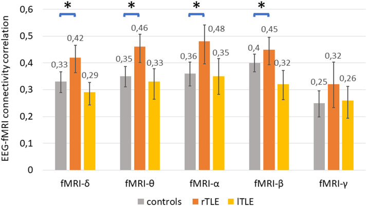
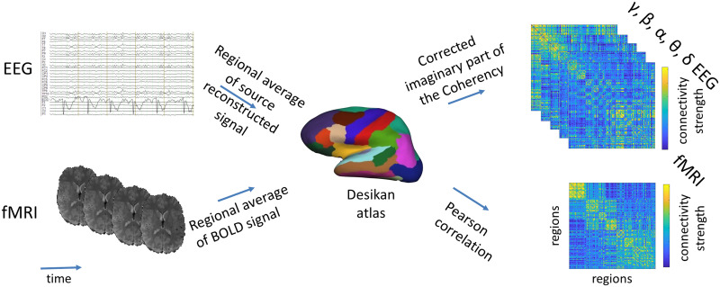
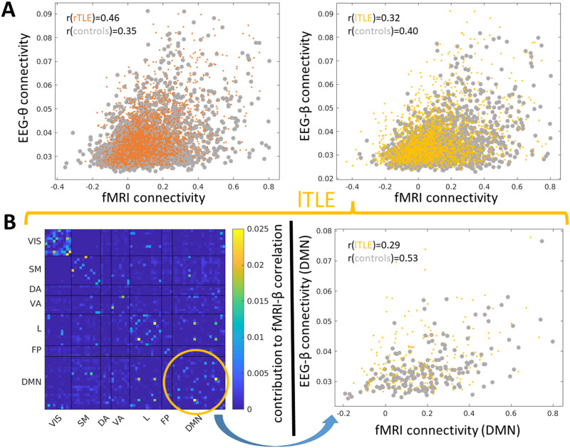
Whole-brain functional connectivity networks (connectomes) have been characterized at different scales in humans using EEG and fMRI. Multimodal epileptic networks have also been investigated, but the relationship between EEG and fMRI defined networks on a whole-brain scale is unclear. A unified multimodal connectome description, mapping healthy and pathological networks would close this knowledge gap. Here, we characterize the spatial correlation between the EEG and fMRI connectomes in right and left temporal lobe epilepsy (rTLE/lTLE). From two centers, we acquired resting-state concurrent EEG-fMRI of 35 healthy controls and 34 TLE patients. EEG-fMRI data was projected into the Desikan brain atlas, and functional connectomes from both modalities were correlated. EEG and fMRI connectomes were moderately correlated. This correlation was increased in rTLE when compared to controls for EEG-delta/theta/alpha/beta. Conversely, multimodal correlation in lTLE was decreased in respect to controls for EEG-beta. While the alteration was global in rTLE, in lTLE it was locally linked to the default mode network. The increased multimodal correlation in rTLE and decreased correlation in lTLE suggests a modality-specific lateralized differential reorganization in TLE, which needs to be considered when comparing results from different modalities. Each modality provides distinct information, highlighting the benefit of multimodal assessment in epilepsy.


 分享
分享
 求助内容:
求助内容: 应助结果提醒方式:
 扫码关注我们
