Shannon Handley, Ayad G. Anwer, Aline Knab, Akanksha Bhargava, Ewa M. Goldys
{"title":"AutoMitoNetwork:用于分析自发荧光图像中线粒体网络的软件,可实现无标记细胞分类。","authors":"Shannon Handley, Ayad G. Anwer, Aline Knab, Akanksha Bhargava, Ewa M. Goldys","doi":"10.1002/cyto.a.24889","DOIUrl":null,"url":null,"abstract":"<p>High-resolution mitochondria imaging in combination with image analysis tools have significantly advanced our understanding of cellular function in health and disease. However, most image analysis tools for mitochondrial studies have been designed to work with fluorescently labeled images only. Additionally, efforts to integrate features describing mitochondrial networks with machine learning techniques for the differentiation of cell types have been limited. Herein, we present AutoMitoNetwork software for image-based assessment of mitochondrial networks in label-free autofluorescence images using a range of interpretable morphological, intensity, and textural features. To demonstrate its utility, we characterized unstained mitochondrial networks in healthy retinal cells and in retinal cells exposed to two types of treatments: rotenone, which directly inhibited mitochondrial respiration and ATP production, and iodoacetic acid, which had a milder impact on mitochondrial networks via the inhibition of anaerobic glycolysis. For both cases, our multi-dimensional feature analysis combined with a support vector machine classifier distinguished between healthy cells and those treated with rotenone or iodoacetic acid. Subtle changes in morphological features were measured including increased fragmentation in the treated retinal cells, pointing to an association with metabolic mechanisms. AutoMitoNetwork opens new options for image-based machine learning in label-free imaging, diagnostics, and mitochondrial disease drug development.</p>","PeriodicalId":11068,"journal":{"name":"Cytometry Part A","volume":"105 9","pages":"688-703"},"PeriodicalIF":2.9000,"publicationDate":"2024-07-30","publicationTypes":"Journal Article","fieldsOfStudy":null,"isOpenAccess":false,"openAccessPdf":"https://onlinelibrary.wiley.com/doi/epdf/10.1002/cyto.a.24889","citationCount":"0","resultStr":"{\"title\":\"AutoMitoNetwork: Software for analyzing mitochondrial networks in autofluorescence images to enable label-free cell classification\",\"authors\":\"Shannon Handley, Ayad G. Anwer, Aline Knab, Akanksha Bhargava, Ewa M. Goldys\",\"doi\":\"10.1002/cyto.a.24889\",\"DOIUrl\":null,\"url\":null,\"abstract\":\"<p>High-resolution mitochondria imaging in combination with image analysis tools have significantly advanced our understanding of cellular function in health and disease. However, most image analysis tools for mitochondrial studies have been designed to work with fluorescently labeled images only. Additionally, efforts to integrate features describing mitochondrial networks with machine learning techniques for the differentiation of cell types have been limited. Herein, we present AutoMitoNetwork software for image-based assessment of mitochondrial networks in label-free autofluorescence images using a range of interpretable morphological, intensity, and textural features. To demonstrate its utility, we characterized unstained mitochondrial networks in healthy retinal cells and in retinal cells exposed to two types of treatments: rotenone, which directly inhibited mitochondrial respiration and ATP production, and iodoacetic acid, which had a milder impact on mitochondrial networks via the inhibition of anaerobic glycolysis. For both cases, our multi-dimensional feature analysis combined with a support vector machine classifier distinguished between healthy cells and those treated with rotenone or iodoacetic acid. Subtle changes in morphological features were measured including increased fragmentation in the treated retinal cells, pointing to an association with metabolic mechanisms. AutoMitoNetwork opens new options for image-based machine learning in label-free imaging, diagnostics, and mitochondrial disease drug development.</p>\",\"PeriodicalId\":11068,\"journal\":{\"name\":\"Cytometry Part A\",\"volume\":\"105 9\",\"pages\":\"688-703\"},\"PeriodicalIF\":2.9000,\"publicationDate\":\"2024-07-30\",\"publicationTypes\":\"Journal Article\",\"fieldsOfStudy\":null,\"isOpenAccess\":false,\"openAccessPdf\":\"https://onlinelibrary.wiley.com/doi/epdf/10.1002/cyto.a.24889\",\"citationCount\":\"0\",\"resultStr\":null,\"platform\":\"Semanticscholar\",\"paperid\":null,\"PeriodicalName\":\"Cytometry Part A\",\"FirstCategoryId\":\"99\",\"ListUrlMain\":\"https://onlinelibrary.wiley.com/doi/10.1002/cyto.a.24889\",\"RegionNum\":4,\"RegionCategory\":\"生物学\",\"ArticlePicture\":[],\"TitleCN\":null,\"AbstractTextCN\":null,\"PMCID\":null,\"EPubDate\":\"\",\"PubModel\":\"\",\"JCR\":\"Q3\",\"JCRName\":\"BIOCHEMICAL RESEARCH METHODS\",\"Score\":null,\"Total\":0}","platform":"Semanticscholar","paperid":null,"PeriodicalName":"Cytometry Part A","FirstCategoryId":"99","ListUrlMain":"https://onlinelibrary.wiley.com/doi/10.1002/cyto.a.24889","RegionNum":4,"RegionCategory":"生物学","ArticlePicture":[],"TitleCN":null,"AbstractTextCN":null,"PMCID":null,"EPubDate":"","PubModel":"","JCR":"Q3","JCRName":"BIOCHEMICAL RESEARCH METHODS","Score":null,"Total":0}
AutoMitoNetwork: Software for analyzing mitochondrial networks in autofluorescence images to enable label-free cell classification
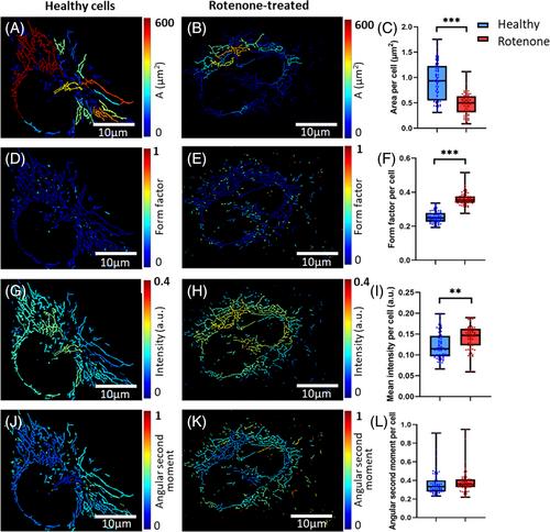
High-resolution mitochondria imaging in combination with image analysis tools have significantly advanced our understanding of cellular function in health and disease. However, most image analysis tools for mitochondrial studies have been designed to work with fluorescently labeled images only. Additionally, efforts to integrate features describing mitochondrial networks with machine learning techniques for the differentiation of cell types have been limited. Herein, we present AutoMitoNetwork software for image-based assessment of mitochondrial networks in label-free autofluorescence images using a range of interpretable morphological, intensity, and textural features. To demonstrate its utility, we characterized unstained mitochondrial networks in healthy retinal cells and in retinal cells exposed to two types of treatments: rotenone, which directly inhibited mitochondrial respiration and ATP production, and iodoacetic acid, which had a milder impact on mitochondrial networks via the inhibition of anaerobic glycolysis. For both cases, our multi-dimensional feature analysis combined with a support vector machine classifier distinguished between healthy cells and those treated with rotenone or iodoacetic acid. Subtle changes in morphological features were measured including increased fragmentation in the treated retinal cells, pointing to an association with metabolic mechanisms. AutoMitoNetwork opens new options for image-based machine learning in label-free imaging, diagnostics, and mitochondrial disease drug development.
期刊介绍:
Cytometry Part A, the journal of quantitative single-cell analysis, features original research reports and reviews of innovative scientific studies employing quantitative single-cell measurement, separation, manipulation, and modeling techniques, as well as original articles on mechanisms of molecular and cellular functions obtained by cytometry techniques.
The journal welcomes submissions from multiple research fields that fully embrace the study of the cytome:
Biomedical Instrumentation Engineering
Biophotonics
Bioinformatics
Cell Biology
Computational Biology
Data Science
Immunology
Parasitology
Microbiology
Neuroscience
Cancer
Stem Cells
Tissue Regeneration.


 分享
分享
 求助内容:
求助内容: 应助结果提醒方式:
 扫码关注我们
