{"title":"心肌细胞外泌体 miR-15a-5p 促进心肌纤维化","authors":"Feng Cao, Zhe Li, Wenmao Ding, Chuan Qv, Hongyi Zhao","doi":"10.1007/s11010-024-05080-3","DOIUrl":null,"url":null,"abstract":"<p><p>The emergence of myofibroblasts is a key step in myocardial fibrosis, but the trigger for the transformation of cardiac fibroblasts into myofibroblasts remains not entirely clear. Exosomes play a key role between cardiomyocytes and cardiac fibroblasts. Here, we not only investigated the relationship between exosomes derived from angiotensin (Ang)-II-treated cardiomyocytes and cardiac fibroblasts, the underlying mechanisms were also explored. Ang-II-treated C57 male mice and mouse cardiac fibroblasts were employed for in vivo and in vitro experiments, respectively. Transmission electron microscopy nanoparticle tracking analysis, and western blot of CD9, CD63, CD81 were performed to identify exosomes; QRT-PCR was performed to detect miR-15a-5p expression; luciferase reporter assay was employed to determine the interaction between miR-15a-5p and dyrk2; western blot was performed to examine the protein levels of fibrosis markers; Counting Kit-8 was performed to determine cell viability; HE and Masson staining were performed to assess the pathological changes of myocardial tissues. MiR-15a-5p expression was found up-regulated in serum of myocardial fibrosis patients, serum and myocardial tissues of Ang-II-treated mice, and Ang-II-treated cardiomyocytes. Mechanically, exosomes from Ang-II-treated cardiomyocytes shuttled miR-15a-5p to cardiac fibroblasts, where miR-15a-5p dephosphorylated NFAT by targeting dyrk2 to promote cell viability and elevated the protein levels of α-smooth muscle actin, collagen type 1 α1 and collagen type 3 α1, thus promoting myocardial fibrosis. This study identified a novel molecular target for anti-fibrotic therapeutic interventions.</p>","PeriodicalId":18724,"journal":{"name":"Molecular and Cellular Biochemistry","volume":" ","pages":"1701-1713"},"PeriodicalIF":3.7000,"publicationDate":"2025-03-01","publicationTypes":"Journal Article","fieldsOfStudy":null,"isOpenAccess":false,"openAccessPdf":"","citationCount":"0","resultStr":"{\"title\":\"Exosomal miR-15a-5p from cardiomyocytes promotes myocardial fibrosis.\",\"authors\":\"Feng Cao, Zhe Li, Wenmao Ding, Chuan Qv, Hongyi Zhao\",\"doi\":\"10.1007/s11010-024-05080-3\",\"DOIUrl\":null,\"url\":null,\"abstract\":\"<p><p>The emergence of myofibroblasts is a key step in myocardial fibrosis, but the trigger for the transformation of cardiac fibroblasts into myofibroblasts remains not entirely clear. Exosomes play a key role between cardiomyocytes and cardiac fibroblasts. Here, we not only investigated the relationship between exosomes derived from angiotensin (Ang)-II-treated cardiomyocytes and cardiac fibroblasts, the underlying mechanisms were also explored. Ang-II-treated C57 male mice and mouse cardiac fibroblasts were employed for in vivo and in vitro experiments, respectively. Transmission electron microscopy nanoparticle tracking analysis, and western blot of CD9, CD63, CD81 were performed to identify exosomes; QRT-PCR was performed to detect miR-15a-5p expression; luciferase reporter assay was employed to determine the interaction between miR-15a-5p and dyrk2; western blot was performed to examine the protein levels of fibrosis markers; Counting Kit-8 was performed to determine cell viability; HE and Masson staining were performed to assess the pathological changes of myocardial tissues. MiR-15a-5p expression was found up-regulated in serum of myocardial fibrosis patients, serum and myocardial tissues of Ang-II-treated mice, and Ang-II-treated cardiomyocytes. Mechanically, exosomes from Ang-II-treated cardiomyocytes shuttled miR-15a-5p to cardiac fibroblasts, where miR-15a-5p dephosphorylated NFAT by targeting dyrk2 to promote cell viability and elevated the protein levels of α-smooth muscle actin, collagen type 1 α1 and collagen type 3 α1, thus promoting myocardial fibrosis. This study identified a novel molecular target for anti-fibrotic therapeutic interventions.</p>\",\"PeriodicalId\":18724,\"journal\":{\"name\":\"Molecular and Cellular Biochemistry\",\"volume\":\" \",\"pages\":\"1701-1713\"},\"PeriodicalIF\":3.7000,\"publicationDate\":\"2025-03-01\",\"publicationTypes\":\"Journal Article\",\"fieldsOfStudy\":null,\"isOpenAccess\":false,\"openAccessPdf\":\"\",\"citationCount\":\"0\",\"resultStr\":null,\"platform\":\"Semanticscholar\",\"paperid\":null,\"PeriodicalName\":\"Molecular and Cellular Biochemistry\",\"FirstCategoryId\":\"99\",\"ListUrlMain\":\"https://doi.org/10.1007/s11010-024-05080-3\",\"RegionNum\":2,\"RegionCategory\":\"生物学\",\"ArticlePicture\":[],\"TitleCN\":null,\"AbstractTextCN\":null,\"PMCID\":null,\"EPubDate\":\"2024/8/7 0:00:00\",\"PubModel\":\"Epub\",\"JCR\":\"Q3\",\"JCRName\":\"CELL BIOLOGY\",\"Score\":null,\"Total\":0}","platform":"Semanticscholar","paperid":null,"PeriodicalName":"Molecular and Cellular Biochemistry","FirstCategoryId":"99","ListUrlMain":"https://doi.org/10.1007/s11010-024-05080-3","RegionNum":2,"RegionCategory":"生物学","ArticlePicture":[],"TitleCN":null,"AbstractTextCN":null,"PMCID":null,"EPubDate":"2024/8/7 0:00:00","PubModel":"Epub","JCR":"Q3","JCRName":"CELL BIOLOGY","Score":null,"Total":0}
Exosomal miR-15a-5p from cardiomyocytes promotes myocardial fibrosis.
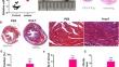
The emergence of myofibroblasts is a key step in myocardial fibrosis, but the trigger for the transformation of cardiac fibroblasts into myofibroblasts remains not entirely clear. Exosomes play a key role between cardiomyocytes and cardiac fibroblasts. Here, we not only investigated the relationship between exosomes derived from angiotensin (Ang)-II-treated cardiomyocytes and cardiac fibroblasts, the underlying mechanisms were also explored. Ang-II-treated C57 male mice and mouse cardiac fibroblasts were employed for in vivo and in vitro experiments, respectively. Transmission electron microscopy nanoparticle tracking analysis, and western blot of CD9, CD63, CD81 were performed to identify exosomes; QRT-PCR was performed to detect miR-15a-5p expression; luciferase reporter assay was employed to determine the interaction between miR-15a-5p and dyrk2; western blot was performed to examine the protein levels of fibrosis markers; Counting Kit-8 was performed to determine cell viability; HE and Masson staining were performed to assess the pathological changes of myocardial tissues. MiR-15a-5p expression was found up-regulated in serum of myocardial fibrosis patients, serum and myocardial tissues of Ang-II-treated mice, and Ang-II-treated cardiomyocytes. Mechanically, exosomes from Ang-II-treated cardiomyocytes shuttled miR-15a-5p to cardiac fibroblasts, where miR-15a-5p dephosphorylated NFAT by targeting dyrk2 to promote cell viability and elevated the protein levels of α-smooth muscle actin, collagen type 1 α1 and collagen type 3 α1, thus promoting myocardial fibrosis. This study identified a novel molecular target for anti-fibrotic therapeutic interventions.
期刊介绍:
Molecular and Cellular Biochemistry: An International Journal for Chemical Biology in Health and Disease publishes original research papers and short communications in all areas of the biochemical sciences, emphasizing novel findings relevant to the biochemical basis of cellular function and disease processes, as well as the mechanics of action of hormones and chemical agents. Coverage includes membrane transport, receptor mechanism, immune response, secretory processes, and cytoskeletal function, as well as biochemical structure-function relationships in the cell.
In addition to the reports of original research, the journal publishes state of the art reviews. Specific subjects covered by Molecular and Cellular Biochemistry include cellular metabolism, cellular pathophysiology, enzymology, ion transport, lipid biochemistry, membrane biochemistry, molecular biology, nuclear structure and function, and protein chemistry.


 分享
分享
 求助内容:
求助内容: 应助结果提醒方式:
 扫码关注我们
