利用小鼠进行多囊肾病研究的机器人超声波和新型胶原分析。

IF 3.2 Q1 UROLOGY & NEPHROLOGY Kidney360 Pub Date : 2024-08-12 DOI:10.34067/KID.0000000000000542
Caroline R Sussman, Heather L Holmes, Alison Stiller, Ka Thao, Adriana V Gregory, Deema Anaam, Ryan Meloche, Yaman Mkhaimer, Harrison H Wells, Luiz D Vasconcelos, Matthew W Urban, Slobodan I Macura, Peter C Harris, Timothy L Kline, Michael F Romero
{"title":"利用小鼠进行多囊肾病研究的机器人超声波和新型胶原分析。","authors":"Caroline R Sussman, Heather L Holmes, Alison Stiller, Ka Thao, Adriana V Gregory, Deema Anaam, Ryan Meloche, Yaman Mkhaimer, Harrison H Wells, Luiz D Vasconcelos, Matthew W Urban, Slobodan I Macura, Peter C Harris, Timothy L Kline, Michael F Romero","doi":"10.34067/KID.0000000000000542","DOIUrl":null,"url":null,"abstract":"<p><strong>Background: </strong>3D imaging and histology are critical tools for assessing polycystic kidney disease ( PKD ) in patients and animal models. Magnetic resonance ( MR ) imaging provides micron resolution, but is time consuming, expensive, and access to equipment and expertise is limiting. Robotic ultrasound ( US ) imaging has lower spatial resolution but is faster, more cost effective, and accessible. Similarly, Picrosirius red ( PSR ) staining and brightfield microscopy is commonly used to assess fibrosis; however, alternative methods have been shown in non-kidney tissues to provide greater sensitivity and more detailed structural characterization.</p><p><strong>Methods: </strong>In this study, we evaluated the utility of robotic US and alternative methods of quantifying PSR staining for PKD research. We compared longitudinal total kidney volume ( TKV ) measurements using US and MR. We additionally compared PSR imaging and quantification using standard brightfield with that by circularly polarized light with hue analysis, and fluorescence imaging analyzed using CT-FIRE software for automatic detection of individual collagen fibers.</p><p><strong>Results: </strong>Increased TKV was detected by US in Pkd1RC/RC vs wild type ( WT ) at timepoints spanning early to established disease. US inter-observer variability was greater but allowed scanning in 2-5 minutes/mouse while MR required 20-30 minutes/mouse. While no change in fibrotic index was detected in this cohort of relatively mild disease using brightfield, polarized light showed fibers skewed thinner in Pkd1RC/RC vs WT. Fluorescence imaging showed a higher density of collagen fibers in Pkd1RC/RC vs WT, and fibers were thinner and curvier with no change in length. Additionally, fiber density was higher in both glomeruli and tubules in Pkd1RC/RC , and glomeruli had a higher fiber density than tubules in Pkd1RC/RC , and trended higher in WT.</p><p><strong>Conclusions: </strong>These studies show robotic ultrasound is a rigorous imaging tool for pre-clinical PKD research. Additionally, they demonstrate the increased sensitivity of polarized and fluorescence analysis of PSR-stained collagen.</p>","PeriodicalId":17882,"journal":{"name":"Kidney360","volume":null,"pages":null},"PeriodicalIF":3.2000,"publicationDate":"2024-08-12","publicationTypes":"Journal Article","fieldsOfStudy":null,"isOpenAccess":false,"openAccessPdf":"","citationCount":"0","resultStr":"{\"title\":\"Robotic Ultrasound and Novel Collagen Analyses for Polycystic Kidney Disease Research Using Mice.\",\"authors\":\"Caroline R Sussman, Heather L Holmes, Alison Stiller, Ka Thao, Adriana V Gregory, Deema Anaam, Ryan Meloche, Yaman Mkhaimer, Harrison H Wells, Luiz D Vasconcelos, Matthew W Urban, Slobodan I Macura, Peter C Harris, Timothy L Kline, Michael F Romero\",\"doi\":\"10.34067/KID.0000000000000542\",\"DOIUrl\":null,\"url\":null,\"abstract\":\"<p><strong>Background: </strong>3D imaging and histology are critical tools for assessing polycystic kidney disease ( PKD ) in patients and animal models. Magnetic resonance ( MR ) imaging provides micron resolution, but is time consuming, expensive, and access to equipment and expertise is limiting. Robotic ultrasound ( US ) imaging has lower spatial resolution but is faster, more cost effective, and accessible. Similarly, Picrosirius red ( PSR ) staining and brightfield microscopy is commonly used to assess fibrosis; however, alternative methods have been shown in non-kidney tissues to provide greater sensitivity and more detailed structural characterization.</p><p><strong>Methods: </strong>In this study, we evaluated the utility of robotic US and alternative methods of quantifying PSR staining for PKD research. We compared longitudinal total kidney volume ( TKV ) measurements using US and MR. We additionally compared PSR imaging and quantification using standard brightfield with that by circularly polarized light with hue analysis, and fluorescence imaging analyzed using CT-FIRE software for automatic detection of individual collagen fibers.</p><p><strong>Results: </strong>Increased TKV was detected by US in Pkd1RC/RC vs wild type ( WT ) at timepoints spanning early to established disease. US inter-observer variability was greater but allowed scanning in 2-5 minutes/mouse while MR required 20-30 minutes/mouse. While no change in fibrotic index was detected in this cohort of relatively mild disease using brightfield, polarized light showed fibers skewed thinner in Pkd1RC/RC vs WT. Fluorescence imaging showed a higher density of collagen fibers in Pkd1RC/RC vs WT, and fibers were thinner and curvier with no change in length. Additionally, fiber density was higher in both glomeruli and tubules in Pkd1RC/RC , and glomeruli had a higher fiber density than tubules in Pkd1RC/RC , and trended higher in WT.</p><p><strong>Conclusions: </strong>These studies show robotic ultrasound is a rigorous imaging tool for pre-clinical PKD research. Additionally, they demonstrate the increased sensitivity of polarized and fluorescence analysis of PSR-stained collagen.</p>\",\"PeriodicalId\":17882,\"journal\":{\"name\":\"Kidney360\",\"volume\":null,\"pages\":null},\"PeriodicalIF\":3.2000,\"publicationDate\":\"2024-08-12\",\"publicationTypes\":\"Journal Article\",\"fieldsOfStudy\":null,\"isOpenAccess\":false,\"openAccessPdf\":\"\",\"citationCount\":\"0\",\"resultStr\":null,\"platform\":\"Semanticscholar\",\"paperid\":null,\"PeriodicalName\":\"Kidney360\",\"FirstCategoryId\":\"1085\",\"ListUrlMain\":\"https://doi.org/10.34067/KID.0000000000000542\",\"RegionNum\":0,\"RegionCategory\":null,\"ArticlePicture\":[],\"TitleCN\":null,\"AbstractTextCN\":null,\"PMCID\":null,\"EPubDate\":\"\",\"PubModel\":\"\",\"JCR\":\"Q1\",\"JCRName\":\"UROLOGY & NEPHROLOGY\",\"Score\":null,\"Total\":0}","platform":"Semanticscholar","paperid":null,"PeriodicalName":"Kidney360","FirstCategoryId":"1085","ListUrlMain":"https://doi.org/10.34067/KID.0000000000000542","RegionNum":0,"RegionCategory":null,"ArticlePicture":[],"TitleCN":null,"AbstractTextCN":null,"PMCID":null,"EPubDate":"","PubModel":"","JCR":"Q1","JCRName":"UROLOGY & NEPHROLOGY","Score":null,"Total":0}
引用次数: 0

摘要

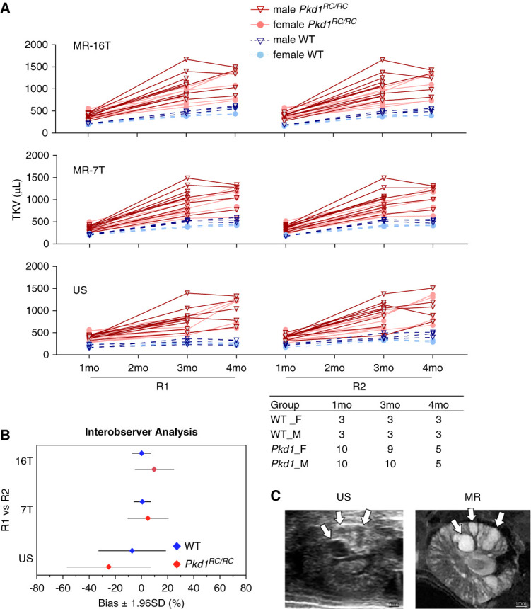
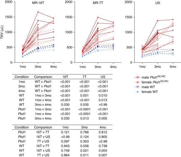
背景:三维成像和组织学是评估患者和动物模型多囊肾病(PKD)的重要工具。磁共振(MR)成像具有微米级分辨率,但耗时长、价格昂贵,而且设备和专业知识的获取受到限制。机器人超声(US)成像的空间分辨率较低,但速度更快、成本效益更高、更容易获得。同样,毕克西里乌斯红(PSR)染色和明视野显微镜通常用于评估纤维化;但在非肾组织中,替代方法已被证明能提供更高的灵敏度和更详细的结构特征:在这项研究中,我们评估了机器人US和其他PSR染色量化方法在PKD研究中的实用性。我们比较了使用 US 和 MR 进行的纵向肾脏总体积 (TKV) 测量。此外,我们还比较了使用标准明视野和圆偏振光进行 PSR 成像和量化的色调分析,以及使用 CT-FIRE 软件自动检测单个胶原纤维的荧光成像分析:结果:在疾病早期到成熟期的时间点上,通过 US 检测到 Pkd1RC/RC 与野生型(WT)的 TKV 增加。US 观察者之间的差异较大,但扫描时间为 2-5 分钟/只小鼠,而 MR 需要 20-30 分钟/只小鼠。在这批病情相对较轻的小鼠中,明视野没有检测到纤维化指数的变化,但偏振光显示 Pkd1RC/RC 与 WT 相比,纤维更细。荧光成像显示,Pkd1RC/RC 与 WT 相比,胶原纤维密度更高,纤维更细、更弯曲,但长度没有变化。此外,Pkd1RC/RC 肾小球和肾小管中的纤维密度均较高,Pkd1RC/RC 肾小球的纤维密度高于肾小管,WT 肾小球的纤维密度也呈上升趋势:这些研究表明,机器人超声是临床前PKD研究的一种严谨的成像工具。结论:这些研究表明,机器人超声是临床前 PKD 研究的一种严谨的成像工具。此外,它们还证明了对 PSR 染色胶原进行偏振和荧光分析可提高灵敏度。
本文章由计算机程序翻译,如有差异,请以英文原文为准。
查看原文
分享 分享
微信好友 朋友圈 QQ好友 复制链接
本刊更多论文
Robotic Ultrasound and Novel Collagen Analyses for Polycystic Kidney Disease Research Using Mice.

Background: 3D imaging and histology are critical tools for assessing polycystic kidney disease ( PKD ) in patients and animal models. Magnetic resonance ( MR ) imaging provides micron resolution, but is time consuming, expensive, and access to equipment and expertise is limiting. Robotic ultrasound ( US ) imaging has lower spatial resolution but is faster, more cost effective, and accessible. Similarly, Picrosirius red ( PSR ) staining and brightfield microscopy is commonly used to assess fibrosis; however, alternative methods have been shown in non-kidney tissues to provide greater sensitivity and more detailed structural characterization.

Methods: In this study, we evaluated the utility of robotic US and alternative methods of quantifying PSR staining for PKD research. We compared longitudinal total kidney volume ( TKV ) measurements using US and MR. We additionally compared PSR imaging and quantification using standard brightfield with that by circularly polarized light with hue analysis, and fluorescence imaging analyzed using CT-FIRE software for automatic detection of individual collagen fibers.

Results: Increased TKV was detected by US in Pkd1RC/RC vs wild type ( WT ) at timepoints spanning early to established disease. US inter-observer variability was greater but allowed scanning in 2-5 minutes/mouse while MR required 20-30 minutes/mouse. While no change in fibrotic index was detected in this cohort of relatively mild disease using brightfield, polarized light showed fibers skewed thinner in Pkd1RC/RC vs WT. Fluorescence imaging showed a higher density of collagen fibers in Pkd1RC/RC vs WT, and fibers were thinner and curvier with no change in length. Additionally, fiber density was higher in both glomeruli and tubules in Pkd1RC/RC , and glomeruli had a higher fiber density than tubules in Pkd1RC/RC , and trended higher in WT.

Conclusions: These studies show robotic ultrasound is a rigorous imaging tool for pre-clinical PKD research. Additionally, they demonstrate the increased sensitivity of polarized and fluorescence analysis of PSR-stained collagen.

求助全文
通过发布文献求助,成功后即可免费获取论文全文。 去求助
来源期刊
Kidney360
Kidney360 UROLOGY & NEPHROLOGY-
CiteScore
3.90
自引率
0.00%
发文量
0
期刊最新文献
The Association of Heart Failure and Edema Events between Patients Initiating SZC or Patiromer. Changes in 24-hour Urine Chemistry in Patients with Nephrolithiasis during Weight Loss with Glucagon-Like Peptide-1 Based Therapies. A Single-Center Retrospective Study to Identify Causes of Sex Differences in the Living Kidney Donor Evaluation Process. Renal Disease and Kidney Transplantation in Hispanic American Persons. Global Perspective of Acute Kidney Injury: Nigeria.
×
引用
GB/T 7714-2015
复制
MLA
复制
APA
复制
导出至
BibTeX EndNote RefMan NoteFirst NoteExpress
×
×
提示
您的信息不完整,为了账户安全,请先补充。
现在去补充
×
提示
您因"违规操作"
具体请查看互助需知
我知道了
×
提示
现在去查看 取消
×
提示
确定
0
微信
客服QQ
Book学术公众号 扫码关注我们
反馈
×
意见反馈
请填写您的意见或建议
请填写您的手机或邮箱
已复制链接
已复制链接
快去分享给好友吧!
我知道了
×
扫码分享
扫码分享
Book学术官方微信
Book学术文献互助
Book学术文献互助群
群 号:481959085
Book学术
文献互助 智能选刊 最新文献 互助须知 联系我们:info@booksci.cn
Book学术提供免费学术资源搜索服务,方便国内外学者检索中英文文献。致力于提供最便捷和优质的服务体验。
Copyright © 2023 Book学术 All rights reserved.
ghs 京公网安备 11010802042870号 京ICP备2023020795号-1