Davide Alaimo, Maria Chiara Terranova, Ettore Palizzolo, Manfredi De Angelis, Vittorio Avella, Giuseppe Paviglianiti, Giuseppe Lo Re, Domenica Matranga, Sergio Salerno
{"title":"评估腕骨年龄的两种不同人工智能(AI)方法与标准 Greulich 和 Pyle 方法的性能比较。","authors":"Davide Alaimo, Maria Chiara Terranova, Ettore Palizzolo, Manfredi De Angelis, Vittorio Avella, Giuseppe Paviglianiti, Giuseppe Lo Re, Domenica Matranga, Sergio Salerno","doi":"10.1007/s11547-024-01871-2","DOIUrl":null,"url":null,"abstract":"<p><strong>Purpose: </strong>Evaluate the agreement between bone age assessments conducted by two distinct machine learning system and standard Greulich and Pyle method.</p><p><strong>Materials and methods: </strong>Carpal radiographs of 225 patients (mean age 8 years and 10 months, SD = 3 years and 1 month) were retrospectively analysed at two separate institutions (October 2018 and May 2022) by both expert radiologists and radiologists in training as well as by two distinct AI software programmes, 16-bit AI<sup>tm</sup> and BoneXpert® in a blinded manner.</p><p><strong>Results: </strong>The bone age range estimated by the 16-bit AI<sup>tm</sup> system in our sample varied between 1 year and 1 month and 15 years and 8 months (mean bone age 9 years and 5 months SD = 3 years and 3 months). BoneXpert® estimated bone age ranged between 8 months and 15 years and 7 months (mean bone age 8 years and 11 months SD = 3 years and 3 months). The average bone age estimated by the Greulich and Pyle method was between 11 months and 14 years, 9 months (mean bone age 8 years and 4 months SD = 3 years and 3 months). Radiologists' assessments using the Greulich and Pyle method were significantly correlated (Pearson's r > 0.80, p < 0.001). There was no statistical difference between BoneXpert® and 16-bit AI<sup>tm</sup> (mean difference = - 0.19, 95%CI = (- 0.45; 0.08)), and the agreement between two measurements varies between - 3.45 (95%CI = (- 3.95; - 3.03) and 3.07 (95%CI - 3.03; 3.57).</p><p><strong>Conclusions: </strong>Both AI methods and GP provide correlated results, although the measurements made by AI were closer to each other compared to the GP method.</p>","PeriodicalId":20817,"journal":{"name":"Radiologia Medica","volume":" ","pages":"1507-1512"},"PeriodicalIF":4.8000,"publicationDate":"2024-10-01","publicationTypes":"Journal Article","fieldsOfStudy":null,"isOpenAccess":false,"openAccessPdf":"https://www.ncbi.nlm.nih.gov/pmc/articles/PMC11480116/pdf/","citationCount":"0","resultStr":"{\"title\":\"Performance of two different artificial intelligence (AI) methods for assessing carpal bone age compared to the standard Greulich and Pyle method.\",\"authors\":\"Davide Alaimo, Maria Chiara Terranova, Ettore Palizzolo, Manfredi De Angelis, Vittorio Avella, Giuseppe Paviglianiti, Giuseppe Lo Re, Domenica Matranga, Sergio Salerno\",\"doi\":\"10.1007/s11547-024-01871-2\",\"DOIUrl\":null,\"url\":null,\"abstract\":\"<p><strong>Purpose: </strong>Evaluate the agreement between bone age assessments conducted by two distinct machine learning system and standard Greulich and Pyle method.</p><p><strong>Materials and methods: </strong>Carpal radiographs of 225 patients (mean age 8 years and 10 months, SD = 3 years and 1 month) were retrospectively analysed at two separate institutions (October 2018 and May 2022) by both expert radiologists and radiologists in training as well as by two distinct AI software programmes, 16-bit AI<sup>tm</sup> and BoneXpert® in a blinded manner.</p><p><strong>Results: </strong>The bone age range estimated by the 16-bit AI<sup>tm</sup> system in our sample varied between 1 year and 1 month and 15 years and 8 months (mean bone age 9 years and 5 months SD = 3 years and 3 months). BoneXpert® estimated bone age ranged between 8 months and 15 years and 7 months (mean bone age 8 years and 11 months SD = 3 years and 3 months). The average bone age estimated by the Greulich and Pyle method was between 11 months and 14 years, 9 months (mean bone age 8 years and 4 months SD = 3 years and 3 months). Radiologists' assessments using the Greulich and Pyle method were significantly correlated (Pearson's r > 0.80, p < 0.001). There was no statistical difference between BoneXpert® and 16-bit AI<sup>tm</sup> (mean difference = - 0.19, 95%CI = (- 0.45; 0.08)), and the agreement between two measurements varies between - 3.45 (95%CI = (- 3.95; - 3.03) and 3.07 (95%CI - 3.03; 3.57).</p><p><strong>Conclusions: </strong>Both AI methods and GP provide correlated results, although the measurements made by AI were closer to each other compared to the GP method.</p>\",\"PeriodicalId\":20817,\"journal\":{\"name\":\"Radiologia Medica\",\"volume\":\" \",\"pages\":\"1507-1512\"},\"PeriodicalIF\":4.8000,\"publicationDate\":\"2024-10-01\",\"publicationTypes\":\"Journal Article\",\"fieldsOfStudy\":null,\"isOpenAccess\":false,\"openAccessPdf\":\"https://www.ncbi.nlm.nih.gov/pmc/articles/PMC11480116/pdf/\",\"citationCount\":\"0\",\"resultStr\":null,\"platform\":\"Semanticscholar\",\"paperid\":null,\"PeriodicalName\":\"Radiologia Medica\",\"FirstCategoryId\":\"3\",\"ListUrlMain\":\"https://doi.org/10.1007/s11547-024-01871-2\",\"RegionNum\":1,\"RegionCategory\":\"医学\",\"ArticlePicture\":[],\"TitleCN\":null,\"AbstractTextCN\":null,\"PMCID\":null,\"EPubDate\":\"2024/8/20 0:00:00\",\"PubModel\":\"Epub\",\"JCR\":\"Q1\",\"JCRName\":\"RADIOLOGY, NUCLEAR MEDICINE & MEDICAL IMAGING\",\"Score\":null,\"Total\":0}","platform":"Semanticscholar","paperid":null,"PeriodicalName":"Radiologia Medica","FirstCategoryId":"3","ListUrlMain":"https://doi.org/10.1007/s11547-024-01871-2","RegionNum":1,"RegionCategory":"医学","ArticlePicture":[],"TitleCN":null,"AbstractTextCN":null,"PMCID":null,"EPubDate":"2024/8/20 0:00:00","PubModel":"Epub","JCR":"Q1","JCRName":"RADIOLOGY, NUCLEAR MEDICINE & MEDICAL IMAGING","Score":null,"Total":0}
Performance of two different artificial intelligence (AI) methods for assessing carpal bone age compared to the standard Greulich and Pyle method.
Purpose: Evaluate the agreement between bone age assessments conducted by two distinct machine learning system and standard Greulich and Pyle method.
Materials and methods: Carpal radiographs of 225 patients (mean age 8 years and 10 months, SD = 3 years and 1 month) were retrospectively analysed at two separate institutions (October 2018 and May 2022) by both expert radiologists and radiologists in training as well as by two distinct AI software programmes, 16-bit AItm and BoneXpert® in a blinded manner.
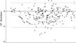
Results: The bone age range estimated by the 16-bit AItm system in our sample varied between 1 year and 1 month and 15 years and 8 months (mean bone age 9 years and 5 months SD = 3 years and 3 months). BoneXpert® estimated bone age ranged between 8 months and 15 years and 7 months (mean bone age 8 years and 11 months SD = 3 years and 3 months). The average bone age estimated by the Greulich and Pyle method was between 11 months and 14 years, 9 months (mean bone age 8 years and 4 months SD = 3 years and 3 months). Radiologists' assessments using the Greulich and Pyle method were significantly correlated (Pearson's r > 0.80, p < 0.001). There was no statistical difference between BoneXpert® and 16-bit AItm (mean difference = - 0.19, 95%CI = (- 0.45; 0.08)), and the agreement between two measurements varies between - 3.45 (95%CI = (- 3.95; - 3.03) and 3.07 (95%CI - 3.03; 3.57).
Conclusions: Both AI methods and GP provide correlated results, although the measurements made by AI were closer to each other compared to the GP method.
期刊介绍:
Felice Perussia founded La radiologia medica in 1914. It is a peer-reviewed journal and serves as the official journal of the Italian Society of Medical and Interventional Radiology (SIRM). The primary purpose of the journal is to disseminate information related to Radiology, especially advancements in diagnostic imaging and related disciplines. La radiologia medica welcomes original research on both fundamental and clinical aspects of modern radiology, with a particular focus on diagnostic and interventional imaging techniques. It also covers topics such as radiotherapy, nuclear medicine, radiobiology, health physics, and artificial intelligence in the context of clinical implications. The journal includes various types of contributions such as original articles, review articles, editorials, short reports, and letters to the editor. With an esteemed Editorial Board and a selection of insightful reports, the journal is an indispensable resource for radiologists and professionals in related fields. Ultimately, La radiologia medica aims to serve as a platform for international collaboration and knowledge sharing within the radiological community.


 分享
分享
 求助内容:
求助内容: 应助结果提醒方式:
 扫码关注我们
