{"title":"利用数字乳腺 X 射线照相术中的 X 射线曝光条件,开发和验证乳腺体积密度推测模型。","authors":"Mika Yamamuro, Yoshiyuki Asai, Takahiro Yamada, Yuichi Kimura, Kazunari Ishii, Yohan Kondo","doi":"10.1007/s11517-024-03186-w","DOIUrl":null,"url":null,"abstract":"<p><p>The use of breast density as a biomarker for breast cancer treatment has not been well established owing to the difficulty in measuring time-series changes in breast density. In this study, we developed a surmising model for breast density using prior mammograms through a multiple regression analysis, enabling a time series analysis of breast density. We acquired 1320 mediolateral oblique view mammograms to construct the surmising model using multiple regression analysis. The dependent variable was the breast density of the mammary gland region segmented by certified radiological technologists, and independent variables included the compressed breast thickness (CBT), exposure current times exposure second (mAs), tube voltage (kV), and patients' age. The coefficient of determination of the surmising model was 0.868. After applying the model, the correlation coefficients of the three groups based on the CBT (thin group, 18-36 mm; standard group, 38-46 mm; and thick group, 48-78 mm) were 0.913, 0.945, and 0.867, respectively, suggesting that the thick breast group had a significantly low correlation coefficient (p = 0.00231). In conclusion, breast density can be accurately surmised using the CBT, mAs, tube voltage, and patients' age, even in the absence of a mammogram image.</p>","PeriodicalId":49840,"journal":{"name":"Medical & Biological Engineering & Computing","volume":" ","pages":"169-179"},"PeriodicalIF":2.6000,"publicationDate":"2025-01-01","publicationTypes":"Journal Article","fieldsOfStudy":null,"isOpenAccess":false,"openAccessPdf":"","citationCount":"0","resultStr":"{\"title\":\"Development and validation of the surmising model for volumetric breast density using X-ray exposure conditions in digital mammography.\",\"authors\":\"Mika Yamamuro, Yoshiyuki Asai, Takahiro Yamada, Yuichi Kimura, Kazunari Ishii, Yohan Kondo\",\"doi\":\"10.1007/s11517-024-03186-w\",\"DOIUrl\":null,\"url\":null,\"abstract\":\"<p><p>The use of breast density as a biomarker for breast cancer treatment has not been well established owing to the difficulty in measuring time-series changes in breast density. In this study, we developed a surmising model for breast density using prior mammograms through a multiple regression analysis, enabling a time series analysis of breast density. We acquired 1320 mediolateral oblique view mammograms to construct the surmising model using multiple regression analysis. The dependent variable was the breast density of the mammary gland region segmented by certified radiological technologists, and independent variables included the compressed breast thickness (CBT), exposure current times exposure second (mAs), tube voltage (kV), and patients' age. The coefficient of determination of the surmising model was 0.868. After applying the model, the correlation coefficients of the three groups based on the CBT (thin group, 18-36 mm; standard group, 38-46 mm; and thick group, 48-78 mm) were 0.913, 0.945, and 0.867, respectively, suggesting that the thick breast group had a significantly low correlation coefficient (p = 0.00231). In conclusion, breast density can be accurately surmised using the CBT, mAs, tube voltage, and patients' age, even in the absence of a mammogram image.</p>\",\"PeriodicalId\":49840,\"journal\":{\"name\":\"Medical & Biological Engineering & Computing\",\"volume\":\" \",\"pages\":\"169-179\"},\"PeriodicalIF\":2.6000,\"publicationDate\":\"2025-01-01\",\"publicationTypes\":\"Journal Article\",\"fieldsOfStudy\":null,\"isOpenAccess\":false,\"openAccessPdf\":\"\",\"citationCount\":\"0\",\"resultStr\":null,\"platform\":\"Semanticscholar\",\"paperid\":null,\"PeriodicalName\":\"Medical & Biological Engineering & Computing\",\"FirstCategoryId\":\"5\",\"ListUrlMain\":\"https://doi.org/10.1007/s11517-024-03186-w\",\"RegionNum\":4,\"RegionCategory\":\"医学\",\"ArticlePicture\":[],\"TitleCN\":null,\"AbstractTextCN\":null,\"PMCID\":null,\"EPubDate\":\"2024/9/2 0:00:00\",\"PubModel\":\"Epub\",\"JCR\":\"Q2\",\"JCRName\":\"COMPUTER SCIENCE, INTERDISCIPLINARY APPLICATIONS\",\"Score\":null,\"Total\":0}","platform":"Semanticscholar","paperid":null,"PeriodicalName":"Medical & Biological Engineering & Computing","FirstCategoryId":"5","ListUrlMain":"https://doi.org/10.1007/s11517-024-03186-w","RegionNum":4,"RegionCategory":"医学","ArticlePicture":[],"TitleCN":null,"AbstractTextCN":null,"PMCID":null,"EPubDate":"2024/9/2 0:00:00","PubModel":"Epub","JCR":"Q2","JCRName":"COMPUTER SCIENCE, INTERDISCIPLINARY APPLICATIONS","Score":null,"Total":0}
引用次数: 0
摘要
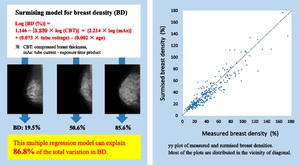
由于难以测量乳腺密度的时间序列变化,因此将乳腺密度作为乳腺癌治疗的生物标志物尚未得到很好的证实。在这项研究中,我们通过多元回归分析,利用先前的乳房X光照片建立了乳房密度推测模型,从而实现了乳房密度的时间序列分析。我们采集了 1320 张内侧斜视乳房 X 光照片,利用多元回归分析建立了推测模型。因变量为经认证的放射技师分割的乳腺区域的乳腺密度,自变量包括压缩乳腺厚度(CBT)、曝光电流乘以曝光秒(mAs)、管电压(kV)和患者年龄。推测模型的决定系数为 0.868。应用该模型后,基于 CBT 的三组(薄组,18-36 毫米;标准组,38-46 毫米;厚组,48-78 毫米)的相关系数分别为 0.913、0.945 和 0.867,表明厚乳房组的相关系数明显较低(p = 0.00231)。总之,即使没有乳房 X 光图像,也可以通过 CBT、mAs、管电压和患者年龄准确推测乳房密度。
Development and validation of the surmising model for volumetric breast density using X-ray exposure conditions in digital mammography.
The use of breast density as a biomarker for breast cancer treatment has not been well established owing to the difficulty in measuring time-series changes in breast density. In this study, we developed a surmising model for breast density using prior mammograms through a multiple regression analysis, enabling a time series analysis of breast density. We acquired 1320 mediolateral oblique view mammograms to construct the surmising model using multiple regression analysis. The dependent variable was the breast density of the mammary gland region segmented by certified radiological technologists, and independent variables included the compressed breast thickness (CBT), exposure current times exposure second (mAs), tube voltage (kV), and patients' age. The coefficient of determination of the surmising model was 0.868. After applying the model, the correlation coefficients of the three groups based on the CBT (thin group, 18-36 mm; standard group, 38-46 mm; and thick group, 48-78 mm) were 0.913, 0.945, and 0.867, respectively, suggesting that the thick breast group had a significantly low correlation coefficient (p = 0.00231). In conclusion, breast density can be accurately surmised using the CBT, mAs, tube voltage, and patients' age, even in the absence of a mammogram image.
期刊介绍:
Founded in 1963, Medical & Biological Engineering & Computing (MBEC) continues to serve the biomedical engineering community, covering the entire spectrum of biomedical and clinical engineering. The journal presents exciting and vital experimental and theoretical developments in biomedical science and technology, and reports on advances in computer-based methodologies in these multidisciplinary subjects. The journal also incorporates new and evolving technologies including cellular engineering and molecular imaging.
MBEC publishes original research articles as well as reviews and technical notes. Its Rapid Communications category focuses on material of immediate value to the readership, while the Controversies section provides a forum to exchange views on selected issues, stimulating a vigorous and informed debate in this exciting and high profile field.
MBEC is an official journal of the International Federation of Medical and Biological Engineering (IFMBE).


 分享
分享
 求助内容:
求助内容: 应助结果提醒方式:
 扫码关注我们
