{"title":"使用放射性标记的亚硝基探针检测脑缺血/再灌注损伤产生的脂质自由基。","authors":"Risa Azuma, Toshihide Yamasaki, Kohei Sano, Takahiro Mukai","doi":"10.1016/j.freeradbiomed.2024.09.025","DOIUrl":null,"url":null,"abstract":"<div><div>Reactive oxygen species generated via reperfusion cause lipid damage and induce lipid peroxidation, leading to cerebral ischemia/reperfusion injury and exacerbation of cerebral infarction. Lipid radicals are key molecules generated during lipid peroxidation. Therefore, understanding the spatiotemporal behavior of lipid radicals is important to improve the therapeutic outcomes of cerebral infarction. However, the behaviors of lipid radicals in the brain remain unclear. In this study, we aimed to evaluate the distribution of radioactivity in a transient middle cerebral artery occlusion (tMCAO) model using lipid radical detection probe [<sup>125</sup>I]<strong>1</strong> to assess the behaviors of lipid radicals after cerebral ischemia/reperfusion. The tMCAO model administered [<sup>125</sup>I]<strong>1</strong> exhibited significant differences in the timing and location of radioactivity accumulation between the ischemic and non-ischemic regions. Liquid chromatography/mass spectrometry analysis identified the lipid radical adducts formed by the reaction of <strong>1</strong> with the lipid radicals generated after reperfusion. More adducts were detected in the ischemic region samples than in the non-ischemic region samples. Therefore, <strong>1</strong> successfully detected the lipid radicals generated after cerebral ischemia/reperfusion. Overall, this study demonstrates the potential of nuclear medical imaging using radiolabeled <strong>1</strong> to detect the lipid radicals generated after cerebral ischemia/reperfusion. Our approach can aid in the development of new therapeutic agents scavenging lipid radicals after cerebral reperfusion by facilitating the determination of therapeutic efficacy and optimal administration period.</div></div>","PeriodicalId":12407,"journal":{"name":"Free Radical Biology and Medicine","volume":"224 ","pages":"Pages 678-684"},"PeriodicalIF":8.2000,"publicationDate":"2024-11-01","publicationTypes":"Journal Article","fieldsOfStudy":null,"isOpenAccess":false,"openAccessPdf":"","citationCount":"0","resultStr":"{\"title\":\"Detection of lipid radicals generated via cerebral ischemia/reperfusion injury using a radiolabeled nitroxide probe\",\"authors\":\"Risa Azuma, Toshihide Yamasaki, Kohei Sano, Takahiro Mukai\",\"doi\":\"10.1016/j.freeradbiomed.2024.09.025\",\"DOIUrl\":null,\"url\":null,\"abstract\":\"<div><div>Reactive oxygen species generated via reperfusion cause lipid damage and induce lipid peroxidation, leading to cerebral ischemia/reperfusion injury and exacerbation of cerebral infarction. Lipid radicals are key molecules generated during lipid peroxidation. Therefore, understanding the spatiotemporal behavior of lipid radicals is important to improve the therapeutic outcomes of cerebral infarction. However, the behaviors of lipid radicals in the brain remain unclear. In this study, we aimed to evaluate the distribution of radioactivity in a transient middle cerebral artery occlusion (tMCAO) model using lipid radical detection probe [<sup>125</sup>I]<strong>1</strong> to assess the behaviors of lipid radicals after cerebral ischemia/reperfusion. The tMCAO model administered [<sup>125</sup>I]<strong>1</strong> exhibited significant differences in the timing and location of radioactivity accumulation between the ischemic and non-ischemic regions. Liquid chromatography/mass spectrometry analysis identified the lipid radical adducts formed by the reaction of <strong>1</strong> with the lipid radicals generated after reperfusion. More adducts were detected in the ischemic region samples than in the non-ischemic region samples. Therefore, <strong>1</strong> successfully detected the lipid radicals generated after cerebral ischemia/reperfusion. Overall, this study demonstrates the potential of nuclear medical imaging using radiolabeled <strong>1</strong> to detect the lipid radicals generated after cerebral ischemia/reperfusion. Our approach can aid in the development of new therapeutic agents scavenging lipid radicals after cerebral reperfusion by facilitating the determination of therapeutic efficacy and optimal administration period.</div></div>\",\"PeriodicalId\":12407,\"journal\":{\"name\":\"Free Radical Biology and Medicine\",\"volume\":\"224 \",\"pages\":\"Pages 678-684\"},\"PeriodicalIF\":8.2000,\"publicationDate\":\"2024-11-01\",\"publicationTypes\":\"Journal Article\",\"fieldsOfStudy\":null,\"isOpenAccess\":false,\"openAccessPdf\":\"\",\"citationCount\":\"0\",\"resultStr\":null,\"platform\":\"Semanticscholar\",\"paperid\":null,\"PeriodicalName\":\"Free Radical Biology and Medicine\",\"FirstCategoryId\":\"3\",\"ListUrlMain\":\"https://www.sciencedirect.com/science/article/pii/S0891584924006725\",\"RegionNum\":2,\"RegionCategory\":\"生物学\",\"ArticlePicture\":[],\"TitleCN\":null,\"AbstractTextCN\":null,\"PMCID\":null,\"EPubDate\":\"2024/9/19 0:00:00\",\"PubModel\":\"Epub\",\"JCR\":\"Q1\",\"JCRName\":\"BIOCHEMISTRY & MOLECULAR BIOLOGY\",\"Score\":null,\"Total\":0}","platform":"Semanticscholar","paperid":null,"PeriodicalName":"Free Radical Biology and Medicine","FirstCategoryId":"3","ListUrlMain":"https://www.sciencedirect.com/science/article/pii/S0891584924006725","RegionNum":2,"RegionCategory":"生物学","ArticlePicture":[],"TitleCN":null,"AbstractTextCN":null,"PMCID":null,"EPubDate":"2024/9/19 0:00:00","PubModel":"Epub","JCR":"Q1","JCRName":"BIOCHEMISTRY & MOLECULAR BIOLOGY","Score":null,"Total":0}
Detection of lipid radicals generated via cerebral ischemia/reperfusion injury using a radiolabeled nitroxide probe
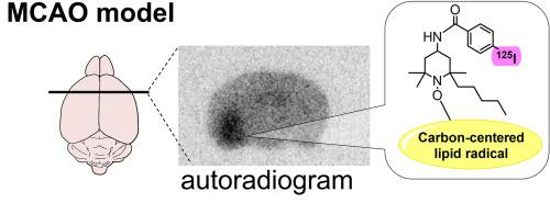
Reactive oxygen species generated via reperfusion cause lipid damage and induce lipid peroxidation, leading to cerebral ischemia/reperfusion injury and exacerbation of cerebral infarction. Lipid radicals are key molecules generated during lipid peroxidation. Therefore, understanding the spatiotemporal behavior of lipid radicals is important to improve the therapeutic outcomes of cerebral infarction. However, the behaviors of lipid radicals in the brain remain unclear. In this study, we aimed to evaluate the distribution of radioactivity in a transient middle cerebral artery occlusion (tMCAO) model using lipid radical detection probe [125I]1 to assess the behaviors of lipid radicals after cerebral ischemia/reperfusion. The tMCAO model administered [125I]1 exhibited significant differences in the timing and location of radioactivity accumulation between the ischemic and non-ischemic regions. Liquid chromatography/mass spectrometry analysis identified the lipid radical adducts formed by the reaction of 1 with the lipid radicals generated after reperfusion. More adducts were detected in the ischemic region samples than in the non-ischemic region samples. Therefore, 1 successfully detected the lipid radicals generated after cerebral ischemia/reperfusion. Overall, this study demonstrates the potential of nuclear medical imaging using radiolabeled 1 to detect the lipid radicals generated after cerebral ischemia/reperfusion. Our approach can aid in the development of new therapeutic agents scavenging lipid radicals after cerebral reperfusion by facilitating the determination of therapeutic efficacy and optimal administration period.
期刊介绍:
Free Radical Biology and Medicine is a leading journal in the field of redox biology, which is the study of the role of reactive oxygen species (ROS) and other oxidizing agents in biological systems. The journal serves as a premier forum for publishing innovative and groundbreaking research that explores the redox biology of health and disease, covering a wide range of topics and disciplines. Free Radical Biology and Medicine also commissions Special Issues that highlight recent advances in both basic and clinical research, with a particular emphasis on the mechanisms underlying altered metabolism and redox signaling. These Special Issues aim to provide a focused platform for the latest research in the field, fostering collaboration and knowledge exchange among researchers and clinicians.


 分享
分享
 求助内容:
求助内容: 应助结果提醒方式:
 扫码关注我们
