Yuding Luo, Gangfeng Gu, Yan Li, Bo Zheng, Fanzhou Ren, Junqiu Wang, Chuanli Chen, Zhao Chen, Yingqian Zhang, Bangcheng Zhao, Jian Yang, Jian Wang
{"title":"检测颈动脉支架内再狭窄的新方法","authors":"Yuding Luo, Gangfeng Gu, Yan Li, Bo Zheng, Fanzhou Ren, Junqiu Wang, Chuanli Chen, Zhao Chen, Yingqian Zhang, Bangcheng Zhao, Jian Yang, Jian Wang","doi":"10.1111/jon.13245","DOIUrl":null,"url":null,"abstract":"<div>\n \n \n <section>\n \n <h3> Background and Purpose</h3>\n \n <p>Carotid artery stenosis is a major risk factor for ischemic stroke. Despite carotid artery stenting, in-stent restenosis (ISR) remains challenging. Pigs serve as an ideal ISR model. This study aims to establish a novel porcine model of carotid ISR using open-loop and closed-loop stents and to assess ISR with optical coherence tomography (OCT) and histopathology, comparing incidence and vascular response between stent types.</p>\n </section>\n \n <section>\n \n <h3> Methods</h3>\n \n <p>Twelve adult male Bama miniature pigs underwent carotid stenting with either open-loop or closed-loop stents. The animals received antiplatelet therapy pre- and postimplantation. Postimplantation evaluations at 90 days included carotid digital subtraction angiography (DSA), OCT, histopathological examination, and electron microscopy.</p>\n </section>\n \n <section>\n \n <h3> Results</h3>\n \n <p>Both stent types showed ISR as detected by OCT and DSA. OCT revealed comparable neointimal proliferation within stent struts for both types, with no significant differences in stent, lumen, and neointimal dimensions. Histopathological analysis and electron microscopy provided insights into tissue responses and healing processes following stent implantation. No significant difference in ISR incidence was found between the stent types based on a <i>χ</i><sup>2</sup> test (<i>p</i> = .110). OCT and hematoxylin-eosin staining exhibit the highest consistency in evaluating neointimal area.</p>\n </section>\n \n <section>\n \n <h3> Conclusions</h3>\n \n <p>The novel porcine ISR model demonstrated similar ISR outcomes for open-loop and closed-loop stents. OCT proved to be a highly consistent and valuable tool for evaluating stent and arterial conditions, comparable to histopathological findings. However, due to a small sample size, the validity of these preliminary findings requires further investigation to be confirmed.</p>\n </section>\n </div>","PeriodicalId":16399,"journal":{"name":"Journal of Neuroimaging","volume":"34 6","pages":"664-672"},"PeriodicalIF":2.3000,"publicationDate":"2024-10-27","publicationTypes":"Journal Article","fieldsOfStudy":null,"isOpenAccess":false,"openAccessPdf":"https://onlinelibrary.wiley.com/doi/epdf/10.1111/jon.13245","citationCount":"0","resultStr":"{\"title\":\"A novel method to detect carotid artery in-stent restenosis\",\"authors\":\"Yuding Luo, Gangfeng Gu, Yan Li, Bo Zheng, Fanzhou Ren, Junqiu Wang, Chuanli Chen, Zhao Chen, Yingqian Zhang, Bangcheng Zhao, Jian Yang, Jian Wang\",\"doi\":\"10.1111/jon.13245\",\"DOIUrl\":null,\"url\":null,\"abstract\":\"<div>\\n \\n \\n <section>\\n \\n <h3> Background and Purpose</h3>\\n \\n <p>Carotid artery stenosis is a major risk factor for ischemic stroke. Despite carotid artery stenting, in-stent restenosis (ISR) remains challenging. Pigs serve as an ideal ISR model. This study aims to establish a novel porcine model of carotid ISR using open-loop and closed-loop stents and to assess ISR with optical coherence tomography (OCT) and histopathology, comparing incidence and vascular response between stent types.</p>\\n </section>\\n \\n <section>\\n \\n <h3> Methods</h3>\\n \\n <p>Twelve adult male Bama miniature pigs underwent carotid stenting with either open-loop or closed-loop stents. The animals received antiplatelet therapy pre- and postimplantation. Postimplantation evaluations at 90 days included carotid digital subtraction angiography (DSA), OCT, histopathological examination, and electron microscopy.</p>\\n </section>\\n \\n <section>\\n \\n <h3> Results</h3>\\n \\n <p>Both stent types showed ISR as detected by OCT and DSA. OCT revealed comparable neointimal proliferation within stent struts for both types, with no significant differences in stent, lumen, and neointimal dimensions. Histopathological analysis and electron microscopy provided insights into tissue responses and healing processes following stent implantation. No significant difference in ISR incidence was found between the stent types based on a <i>χ</i><sup>2</sup> test (<i>p</i> = .110). OCT and hematoxylin-eosin staining exhibit the highest consistency in evaluating neointimal area.</p>\\n </section>\\n \\n <section>\\n \\n <h3> Conclusions</h3>\\n \\n <p>The novel porcine ISR model demonstrated similar ISR outcomes for open-loop and closed-loop stents. OCT proved to be a highly consistent and valuable tool for evaluating stent and arterial conditions, comparable to histopathological findings. However, due to a small sample size, the validity of these preliminary findings requires further investigation to be confirmed.</p>\\n </section>\\n </div>\",\"PeriodicalId\":16399,\"journal\":{\"name\":\"Journal of Neuroimaging\",\"volume\":\"34 6\",\"pages\":\"664-672\"},\"PeriodicalIF\":2.3000,\"publicationDate\":\"2024-10-27\",\"publicationTypes\":\"Journal Article\",\"fieldsOfStudy\":null,\"isOpenAccess\":false,\"openAccessPdf\":\"https://onlinelibrary.wiley.com/doi/epdf/10.1111/jon.13245\",\"citationCount\":\"0\",\"resultStr\":null,\"platform\":\"Semanticscholar\",\"paperid\":null,\"PeriodicalName\":\"Journal of Neuroimaging\",\"FirstCategoryId\":\"3\",\"ListUrlMain\":\"https://onlinelibrary.wiley.com/doi/10.1111/jon.13245\",\"RegionNum\":4,\"RegionCategory\":\"医学\",\"ArticlePicture\":[],\"TitleCN\":null,\"AbstractTextCN\":null,\"PMCID\":null,\"EPubDate\":\"\",\"PubModel\":\"\",\"JCR\":\"Q3\",\"JCRName\":\"CLINICAL NEUROLOGY\",\"Score\":null,\"Total\":0}","platform":"Semanticscholar","paperid":null,"PeriodicalName":"Journal of Neuroimaging","FirstCategoryId":"3","ListUrlMain":"https://onlinelibrary.wiley.com/doi/10.1111/jon.13245","RegionNum":4,"RegionCategory":"医学","ArticlePicture":[],"TitleCN":null,"AbstractTextCN":null,"PMCID":null,"EPubDate":"","PubModel":"","JCR":"Q3","JCRName":"CLINICAL NEUROLOGY","Score":null,"Total":0}
A novel method to detect carotid artery in-stent restenosis
Background and Purpose
Carotid artery stenosis is a major risk factor for ischemic stroke. Despite carotid artery stenting, in-stent restenosis (ISR) remains challenging. Pigs serve as an ideal ISR model. This study aims to establish a novel porcine model of carotid ISR using open-loop and closed-loop stents and to assess ISR with optical coherence tomography (OCT) and histopathology, comparing incidence and vascular response between stent types.
Methods
Twelve adult male Bama miniature pigs underwent carotid stenting with either open-loop or closed-loop stents. The animals received antiplatelet therapy pre- and postimplantation. Postimplantation evaluations at 90 days included carotid digital subtraction angiography (DSA), OCT, histopathological examination, and electron microscopy.
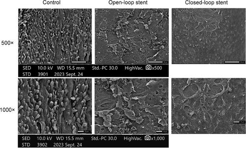
Results
Both stent types showed ISR as detected by OCT and DSA. OCT revealed comparable neointimal proliferation within stent struts for both types, with no significant differences in stent, lumen, and neointimal dimensions. Histopathological analysis and electron microscopy provided insights into tissue responses and healing processes following stent implantation. No significant difference in ISR incidence was found between the stent types based on a χ2 test (p = .110). OCT and hematoxylin-eosin staining exhibit the highest consistency in evaluating neointimal area.
Conclusions
The novel porcine ISR model demonstrated similar ISR outcomes for open-loop and closed-loop stents. OCT proved to be a highly consistent and valuable tool for evaluating stent and arterial conditions, comparable to histopathological findings. However, due to a small sample size, the validity of these preliminary findings requires further investigation to be confirmed.
期刊介绍:
Start reading the Journal of Neuroimaging to learn the latest neurological imaging techniques. The peer-reviewed research is written in a practical clinical context, giving you the information you need on:
MRI
CT
Carotid Ultrasound and TCD
SPECT
PET
Endovascular Surgical Neuroradiology
Functional MRI
Xenon CT
and other new and upcoming neuroscientific modalities.The Journal of Neuroimaging addresses the full spectrum of human nervous system disease, including stroke, neoplasia, degenerating and demyelinating disease, epilepsy, tumors, lesions, infectious disease, cerebral vascular arterial diseases, toxic-metabolic disease, psychoses, dementias, heredo-familial disease, and trauma.Offering original research, review articles, case reports, neuroimaging CPCs, and evaluations of instruments and technology relevant to the nervous system, the Journal of Neuroimaging focuses on useful clinical developments and applications, tested techniques and interpretations, patient care, diagnostics, and therapeutics. Start reading today!


 分享
分享
 求助内容:
求助内容: 应助结果提醒方式:
 扫码关注我们
