Yuankun Zhai, Zhuang Zhou, Xiaojie Xing, Mark Nuzzle, Xinping Zhang
{"title":"颅骨缺损修复中上硬膜和硬膜不同骨和血管类型的形成","authors":"Yuankun Zhai, Zhuang Zhou, Xiaojie Xing, Mark Nuzzle, Xinping Zhang","doi":"10.1038/s41413-024-00379-9","DOIUrl":null,"url":null,"abstract":"<p>The cranial mesenchyme, originating from both neural crest and mesoderm, imparts remarkable regional specificity and complexity to postnatal calvarial tissue. While the distinct embryonic origins of the superior and dura periosteum of the cranial parietal bone have been described, the extent of their respective contributions to bone and vessel formation during adult bone defect repair remains superficially explored. Utilizing transgenic mouse models in conjunction with high-resolution multiphoton laser scanning microscopy (MPLSM), we have separately evaluated bone and vessel formation in the superior and dura periosteum before and after injury, as well as following intermittent treatment of recombinant peptide of human parathyroid hormone (rhPTH), Teriparatide. Our results show that new bone formation along the dura surface is three times greater than that along the superior periosteal surface following injury, regardless of Teriparatide treatment. Targeted deletion of PTH receptor PTH1R via <i>SMA-CreER</i> and <i>Col 1a (2.3)-CreER</i> results in selective reduction of bone formation, suggesting different progenitor cell pools in the adult superior and dura periosteum. Consistently, analyses of microvasculature show higher vessel density and better organized arterial-venous vessel network associated with a 10-fold more osteoblast clusters at dura periosteum as compared to superior periosteum. Intermittent rhPTH treatment further enhances the arterial vessel ratio at dura periosteum and type H vessel formation in cortical bone marrow space. Taken together, our study demonstrates a site-dependent coordinated osteogenic and angiogenic response, which is determined by regional osteogenic progenitor pool as well as the coupling blood vessel network at the site of cranial defect repair.</p>","PeriodicalId":9134,"journal":{"name":"Bone Research","volume":"7 1","pages":""},"PeriodicalIF":15.0000,"publicationDate":"2025-01-13","publicationTypes":"Journal Article","fieldsOfStudy":null,"isOpenAccess":false,"openAccessPdf":"","citationCount":"0","resultStr":"{\"title\":\"Differential bone and vessel type formation at superior and dura periosteum during cranial bone defect repair\",\"authors\":\"Yuankun Zhai, Zhuang Zhou, Xiaojie Xing, Mark Nuzzle, Xinping Zhang\",\"doi\":\"10.1038/s41413-024-00379-9\",\"DOIUrl\":null,\"url\":null,\"abstract\":\"<p>The cranial mesenchyme, originating from both neural crest and mesoderm, imparts remarkable regional specificity and complexity to postnatal calvarial tissue. While the distinct embryonic origins of the superior and dura periosteum of the cranial parietal bone have been described, the extent of their respective contributions to bone and vessel formation during adult bone defect repair remains superficially explored. Utilizing transgenic mouse models in conjunction with high-resolution multiphoton laser scanning microscopy (MPLSM), we have separately evaluated bone and vessel formation in the superior and dura periosteum before and after injury, as well as following intermittent treatment of recombinant peptide of human parathyroid hormone (rhPTH), Teriparatide. Our results show that new bone formation along the dura surface is three times greater than that along the superior periosteal surface following injury, regardless of Teriparatide treatment. Targeted deletion of PTH receptor PTH1R via <i>SMA-CreER</i> and <i>Col 1a (2.3)-CreER</i> results in selective reduction of bone formation, suggesting different progenitor cell pools in the adult superior and dura periosteum. Consistently, analyses of microvasculature show higher vessel density and better organized arterial-venous vessel network associated with a 10-fold more osteoblast clusters at dura periosteum as compared to superior periosteum. Intermittent rhPTH treatment further enhances the arterial vessel ratio at dura periosteum and type H vessel formation in cortical bone marrow space. Taken together, our study demonstrates a site-dependent coordinated osteogenic and angiogenic response, which is determined by regional osteogenic progenitor pool as well as the coupling blood vessel network at the site of cranial defect repair.</p>\",\"PeriodicalId\":9134,\"journal\":{\"name\":\"Bone Research\",\"volume\":\"7 1\",\"pages\":\"\"},\"PeriodicalIF\":15.0000,\"publicationDate\":\"2025-01-13\",\"publicationTypes\":\"Journal Article\",\"fieldsOfStudy\":null,\"isOpenAccess\":false,\"openAccessPdf\":\"\",\"citationCount\":\"0\",\"resultStr\":null,\"platform\":\"Semanticscholar\",\"paperid\":null,\"PeriodicalName\":\"Bone Research\",\"FirstCategoryId\":\"3\",\"ListUrlMain\":\"https://doi.org/10.1038/s41413-024-00379-9\",\"RegionNum\":1,\"RegionCategory\":\"医学\",\"ArticlePicture\":[],\"TitleCN\":null,\"AbstractTextCN\":null,\"PMCID\":null,\"EPubDate\":\"\",\"PubModel\":\"\",\"JCR\":\"Q1\",\"JCRName\":\"CELL & TISSUE ENGINEERING\",\"Score\":null,\"Total\":0}","platform":"Semanticscholar","paperid":null,"PeriodicalName":"Bone Research","FirstCategoryId":"3","ListUrlMain":"https://doi.org/10.1038/s41413-024-00379-9","RegionNum":1,"RegionCategory":"医学","ArticlePicture":[],"TitleCN":null,"AbstractTextCN":null,"PMCID":null,"EPubDate":"","PubModel":"","JCR":"Q1","JCRName":"CELL & TISSUE ENGINEERING","Score":null,"Total":0}
Differential bone and vessel type formation at superior and dura periosteum during cranial bone defect repair
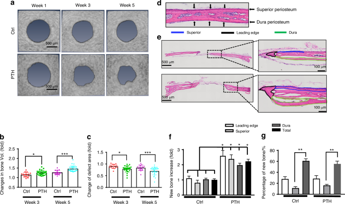
The cranial mesenchyme, originating from both neural crest and mesoderm, imparts remarkable regional specificity and complexity to postnatal calvarial tissue. While the distinct embryonic origins of the superior and dura periosteum of the cranial parietal bone have been described, the extent of their respective contributions to bone and vessel formation during adult bone defect repair remains superficially explored. Utilizing transgenic mouse models in conjunction with high-resolution multiphoton laser scanning microscopy (MPLSM), we have separately evaluated bone and vessel formation in the superior and dura periosteum before and after injury, as well as following intermittent treatment of recombinant peptide of human parathyroid hormone (rhPTH), Teriparatide. Our results show that new bone formation along the dura surface is three times greater than that along the superior periosteal surface following injury, regardless of Teriparatide treatment. Targeted deletion of PTH receptor PTH1R via SMA-CreER and Col 1a (2.3)-CreER results in selective reduction of bone formation, suggesting different progenitor cell pools in the adult superior and dura periosteum. Consistently, analyses of microvasculature show higher vessel density and better organized arterial-venous vessel network associated with a 10-fold more osteoblast clusters at dura periosteum as compared to superior periosteum. Intermittent rhPTH treatment further enhances the arterial vessel ratio at dura periosteum and type H vessel formation in cortical bone marrow space. Taken together, our study demonstrates a site-dependent coordinated osteogenic and angiogenic response, which is determined by regional osteogenic progenitor pool as well as the coupling blood vessel network at the site of cranial defect repair.
期刊介绍:
Established in 2013, Bone Research is a newly-founded English-language periodical that centers on the basic and clinical facets of bone biology, pathophysiology, and regeneration. It is dedicated to championing key findings emerging from both basic investigations and clinical research concerning bone-related topics. The journal's objective is to globally disseminate research in bone-related physiology, pathology, diseases, and treatment, contributing to the advancement of knowledge in this field.


 分享
分享
 求助内容:
求助内容: 应助结果提醒方式:
 扫码关注我们
