Qiao Ma, Kunpeng Ma, Yanli Dong, Yufei Meng, Jun Zhao, Renjie Li, Qinru Bai, Di Wu, Daohua Jiang, Jianyuan Sun, Yan Zhao
{"title":"囊泡型乙酰胆碱转运体VAChT的结合机制及拮抗作用","authors":"Qiao Ma, Kunpeng Ma, Yanli Dong, Yufei Meng, Jun Zhao, Renjie Li, Qinru Bai, Di Wu, Daohua Jiang, Jianyuan Sun, Yan Zhao","doi":"10.1038/s41594-024-01462-9","DOIUrl":null,"url":null,"abstract":"The vesicular acetylcholine transporter (VAChT) has a pivotal role in packaging and transporting acetylcholine for exocytotic release, serving as a vital component of cholinergic neurotransmission. Dysregulation of its function can result in neurological disorders. It also serves as a target for developing radiotracers to quantify cholinergic neuron deficits in neurodegenerative conditions. Here we unveil the cryo-electron microscopy structures of human VAChT in its apo state, the substrate acetylcholine-bound state and the inhibitor vesamicol-bound state. These structures assume a lumen-facing conformation, offering a clear depiction of architecture of VAChT. The acetylcholine-bound structure provides a detailed understanding of how VAChT recognizes its substrate, shedding light on the coupling mechanism of protonation and substrate binding. Meanwhile, the vesamicol-bound structure reveals the binding mode of vesamicol to VAChT, laying the structural foundation for the design of the next generation of radioligands targeting VAChT. Here the authors show structures of the vesicular acetylcholine transporter in its apo state and in complex with the substrate acetylcholine and the inhibitor vesamicol, providing insights into substrate recognition, proton coupling and conformational transition mechanisms.","PeriodicalId":49141,"journal":{"name":"Nature Structural & Molecular Biology","volume":"32 5","pages":"818-827"},"PeriodicalIF":10.1000,"publicationDate":"2025-01-13","publicationTypes":"Journal Article","fieldsOfStudy":null,"isOpenAccess":false,"openAccessPdf":"","citationCount":"0","resultStr":"{\"title\":\"Binding mechanism and antagonism of the vesicular acetylcholine transporter VAChT\",\"authors\":\"Qiao Ma, Kunpeng Ma, Yanli Dong, Yufei Meng, Jun Zhao, Renjie Li, Qinru Bai, Di Wu, Daohua Jiang, Jianyuan Sun, Yan Zhao\",\"doi\":\"10.1038/s41594-024-01462-9\",\"DOIUrl\":null,\"url\":null,\"abstract\":\"The vesicular acetylcholine transporter (VAChT) has a pivotal role in packaging and transporting acetylcholine for exocytotic release, serving as a vital component of cholinergic neurotransmission. Dysregulation of its function can result in neurological disorders. It also serves as a target for developing radiotracers to quantify cholinergic neuron deficits in neurodegenerative conditions. Here we unveil the cryo-electron microscopy structures of human VAChT in its apo state, the substrate acetylcholine-bound state and the inhibitor vesamicol-bound state. These structures assume a lumen-facing conformation, offering a clear depiction of architecture of VAChT. The acetylcholine-bound structure provides a detailed understanding of how VAChT recognizes its substrate, shedding light on the coupling mechanism of protonation and substrate binding. Meanwhile, the vesamicol-bound structure reveals the binding mode of vesamicol to VAChT, laying the structural foundation for the design of the next generation of radioligands targeting VAChT. Here the authors show structures of the vesicular acetylcholine transporter in its apo state and in complex with the substrate acetylcholine and the inhibitor vesamicol, providing insights into substrate recognition, proton coupling and conformational transition mechanisms.\",\"PeriodicalId\":49141,\"journal\":{\"name\":\"Nature Structural & Molecular Biology\",\"volume\":\"32 5\",\"pages\":\"818-827\"},\"PeriodicalIF\":10.1000,\"publicationDate\":\"2025-01-13\",\"publicationTypes\":\"Journal Article\",\"fieldsOfStudy\":null,\"isOpenAccess\":false,\"openAccessPdf\":\"\",\"citationCount\":\"0\",\"resultStr\":null,\"platform\":\"Semanticscholar\",\"paperid\":null,\"PeriodicalName\":\"Nature Structural & Molecular Biology\",\"FirstCategoryId\":\"99\",\"ListUrlMain\":\"https://www.nature.com/articles/s41594-024-01462-9\",\"RegionNum\":1,\"RegionCategory\":\"生物学\",\"ArticlePicture\":[],\"TitleCN\":null,\"AbstractTextCN\":null,\"PMCID\":null,\"EPubDate\":\"\",\"PubModel\":\"\",\"JCR\":\"Q1\",\"JCRName\":\"BIOCHEMISTRY & MOLECULAR BIOLOGY\",\"Score\":null,\"Total\":0}","platform":"Semanticscholar","paperid":null,"PeriodicalName":"Nature Structural & Molecular Biology","FirstCategoryId":"99","ListUrlMain":"https://www.nature.com/articles/s41594-024-01462-9","RegionNum":1,"RegionCategory":"生物学","ArticlePicture":[],"TitleCN":null,"AbstractTextCN":null,"PMCID":null,"EPubDate":"","PubModel":"","JCR":"Q1","JCRName":"BIOCHEMISTRY & MOLECULAR BIOLOGY","Score":null,"Total":0}
Binding mechanism and antagonism of the vesicular acetylcholine transporter VAChT
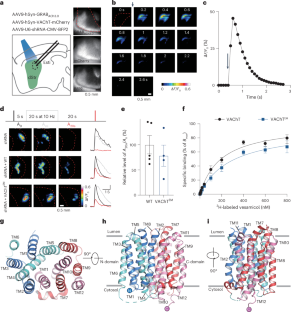
The vesicular acetylcholine transporter (VAChT) has a pivotal role in packaging and transporting acetylcholine for exocytotic release, serving as a vital component of cholinergic neurotransmission. Dysregulation of its function can result in neurological disorders. It also serves as a target for developing radiotracers to quantify cholinergic neuron deficits in neurodegenerative conditions. Here we unveil the cryo-electron microscopy structures of human VAChT in its apo state, the substrate acetylcholine-bound state and the inhibitor vesamicol-bound state. These structures assume a lumen-facing conformation, offering a clear depiction of architecture of VAChT. The acetylcholine-bound structure provides a detailed understanding of how VAChT recognizes its substrate, shedding light on the coupling mechanism of protonation and substrate binding. Meanwhile, the vesamicol-bound structure reveals the binding mode of vesamicol to VAChT, laying the structural foundation for the design of the next generation of radioligands targeting VAChT. Here the authors show structures of the vesicular acetylcholine transporter in its apo state and in complex with the substrate acetylcholine and the inhibitor vesamicol, providing insights into substrate recognition, proton coupling and conformational transition mechanisms.
期刊介绍:
Nature Structural & Molecular Biology is a comprehensive platform that combines structural and molecular research. Our journal focuses on exploring the functional and mechanistic aspects of biological processes, emphasizing how molecular components collaborate to achieve a particular function. While structural data can shed light on these insights, our publication does not require them as a prerequisite.


 分享
分享
 求助内容:
求助内容: 应助结果提醒方式:
 扫码关注我们
