Madeleine Amy Bessen, Christine Diana Gayen, Ryan L O'Hare Doig, Ryan Michael Dorrian, Ryan David Quarrington, Adnan Mulaibrahimovic, Vartan Kurtcuoglu, Angela Catherine Walls, Anna Victoria Leonard, Claire Frances Jones
{"title":"猪外伤性脊髓损伤后的脑脊液动力学和蛛网膜下腔闭塞:利用磁共振成像进行的研究。","authors":"Madeleine Amy Bessen, Christine Diana Gayen, Ryan L O'Hare Doig, Ryan Michael Dorrian, Ryan David Quarrington, Adnan Mulaibrahimovic, Vartan Kurtcuoglu, Angela Catherine Walls, Anna Victoria Leonard, Claire Frances Jones","doi":"10.1186/s12987-024-00595-9","DOIUrl":null,"url":null,"abstract":"<p><strong>Background: </strong>Traumatic spinal cord injury (SCI) causes spinal cord swelling and occlusion of the subarachnoid space (SAS). SAS occlusion can change pulsatile cerebrospinal fluid (CSF) dynamics, which could have acute clinical management implications. This study aimed to characterise SAS occlusion and investigate CSF dynamics over 14 days post-SCI in the pig.</p><p><strong>Methods: </strong>A thoracic contusion SCI was induced in female domestic pigs (22-29 kg) via a weight drop apparatus (N = 5, 10 cm; N = 5, 20 cm). Magnetic resonance imaging (MRI) was performed pre-SCI and 3, 7 and 14 days post-SCI. SAS occlusion length (cranial-caudal), and injury site SAS area (cross-sectional), were measured on T2-weighted MRI. CSF dynamics, specifically peak cranial/caudal mean velocity (cm/s), and the corresponding time to peak (% of cardiac cycle), were measured on cardiac gated, axial phase-contrast MRI obtained at C2/C3, T8/T9, T11/T12 and L1/L2. Linear-mixed effects models, with a significance level of α = 0.05, were developed to assess the effect of: (1) injury group and time point on SAS occlusion measures; and (2), time point and spinal level, adjusted by injury group, on CSF dynamics.</p><p><strong>Results: </strong>For both injury groups, SAS occlusion length decreased from 3 to 7 days post-SCI, and 7 to 14 days post-SCI. The cross-sectional SAS area decreased after SCI, and increased to 14 days post-SCI, in both groups. At all spinal levels, peak cranial/caudal mean velocity and the time to peak caudal mean velocity decreased at day 3 post-SCI. From 3 to 14 days post-SCI, peak caudal mean velocity and the time to peak caudal mean velocity increased towards baseline values, at all spinal levels.</p><p><strong>Conclusions: </strong>Spinal-level specific changes to CSF dynamics, with concurrent changes to SAS occlusion, occurred after SCI in the pig, suggesting that CSF pulsatility and craniospinal compliance were altered in the sub-acute post-traumatic period. These results suggest that PC-MRI derived CSF dynamics may provide a non-invasive method to investigate functional alterations to the spinal intrathecal space following traumatic SCI.</p>","PeriodicalId":12321,"journal":{"name":"Fluids and Barriers of the CNS","volume":"22 1","pages":"6"},"PeriodicalIF":6.2000,"publicationDate":"2025-01-14","publicationTypes":"Journal Article","fieldsOfStudy":null,"isOpenAccess":false,"openAccessPdf":"https://www.ncbi.nlm.nih.gov/pmc/articles/PMC11730158/pdf/","citationCount":"0","resultStr":"{\"title\":\"Cerebrospinal fluid dynamics and subarachnoid space occlusion following traumatic spinal cord injury in the pig: an investigation using magnetic resonance imaging.\",\"authors\":\"Madeleine Amy Bessen, Christine Diana Gayen, Ryan L O'Hare Doig, Ryan Michael Dorrian, Ryan David Quarrington, Adnan Mulaibrahimovic, Vartan Kurtcuoglu, Angela Catherine Walls, Anna Victoria Leonard, Claire Frances Jones\",\"doi\":\"10.1186/s12987-024-00595-9\",\"DOIUrl\":null,\"url\":null,\"abstract\":\"<p><strong>Background: </strong>Traumatic spinal cord injury (SCI) causes spinal cord swelling and occlusion of the subarachnoid space (SAS). SAS occlusion can change pulsatile cerebrospinal fluid (CSF) dynamics, which could have acute clinical management implications. This study aimed to characterise SAS occlusion and investigate CSF dynamics over 14 days post-SCI in the pig.</p><p><strong>Methods: </strong>A thoracic contusion SCI was induced in female domestic pigs (22-29 kg) via a weight drop apparatus (N = 5, 10 cm; N = 5, 20 cm). Magnetic resonance imaging (MRI) was performed pre-SCI and 3, 7 and 14 days post-SCI. SAS occlusion length (cranial-caudal), and injury site SAS area (cross-sectional), were measured on T2-weighted MRI. CSF dynamics, specifically peak cranial/caudal mean velocity (cm/s), and the corresponding time to peak (% of cardiac cycle), were measured on cardiac gated, axial phase-contrast MRI obtained at C2/C3, T8/T9, T11/T12 and L1/L2. Linear-mixed effects models, with a significance level of α = 0.05, were developed to assess the effect of: (1) injury group and time point on SAS occlusion measures; and (2), time point and spinal level, adjusted by injury group, on CSF dynamics.</p><p><strong>Results: </strong>For both injury groups, SAS occlusion length decreased from 3 to 7 days post-SCI, and 7 to 14 days post-SCI. The cross-sectional SAS area decreased after SCI, and increased to 14 days post-SCI, in both groups. At all spinal levels, peak cranial/caudal mean velocity and the time to peak caudal mean velocity decreased at day 3 post-SCI. From 3 to 14 days post-SCI, peak caudal mean velocity and the time to peak caudal mean velocity increased towards baseline values, at all spinal levels.</p><p><strong>Conclusions: </strong>Spinal-level specific changes to CSF dynamics, with concurrent changes to SAS occlusion, occurred after SCI in the pig, suggesting that CSF pulsatility and craniospinal compliance were altered in the sub-acute post-traumatic period. These results suggest that PC-MRI derived CSF dynamics may provide a non-invasive method to investigate functional alterations to the spinal intrathecal space following traumatic SCI.</p>\",\"PeriodicalId\":12321,\"journal\":{\"name\":\"Fluids and Barriers of the CNS\",\"volume\":\"22 1\",\"pages\":\"6\"},\"PeriodicalIF\":6.2000,\"publicationDate\":\"2025-01-14\",\"publicationTypes\":\"Journal Article\",\"fieldsOfStudy\":null,\"isOpenAccess\":false,\"openAccessPdf\":\"https://www.ncbi.nlm.nih.gov/pmc/articles/PMC11730158/pdf/\",\"citationCount\":\"0\",\"resultStr\":null,\"platform\":\"Semanticscholar\",\"paperid\":null,\"PeriodicalName\":\"Fluids and Barriers of the CNS\",\"FirstCategoryId\":\"3\",\"ListUrlMain\":\"https://doi.org/10.1186/s12987-024-00595-9\",\"RegionNum\":1,\"RegionCategory\":\"医学\",\"ArticlePicture\":[],\"TitleCN\":null,\"AbstractTextCN\":null,\"PMCID\":null,\"EPubDate\":\"\",\"PubModel\":\"\",\"JCR\":\"Q1\",\"JCRName\":\"NEUROSCIENCES\",\"Score\":null,\"Total\":0}","platform":"Semanticscholar","paperid":null,"PeriodicalName":"Fluids and Barriers of the CNS","FirstCategoryId":"3","ListUrlMain":"https://doi.org/10.1186/s12987-024-00595-9","RegionNum":1,"RegionCategory":"医学","ArticlePicture":[],"TitleCN":null,"AbstractTextCN":null,"PMCID":null,"EPubDate":"","PubModel":"","JCR":"Q1","JCRName":"NEUROSCIENCES","Score":null,"Total":0}
Cerebrospinal fluid dynamics and subarachnoid space occlusion following traumatic spinal cord injury in the pig: an investigation using magnetic resonance imaging.
Background: Traumatic spinal cord injury (SCI) causes spinal cord swelling and occlusion of the subarachnoid space (SAS). SAS occlusion can change pulsatile cerebrospinal fluid (CSF) dynamics, which could have acute clinical management implications. This study aimed to characterise SAS occlusion and investigate CSF dynamics over 14 days post-SCI in the pig.
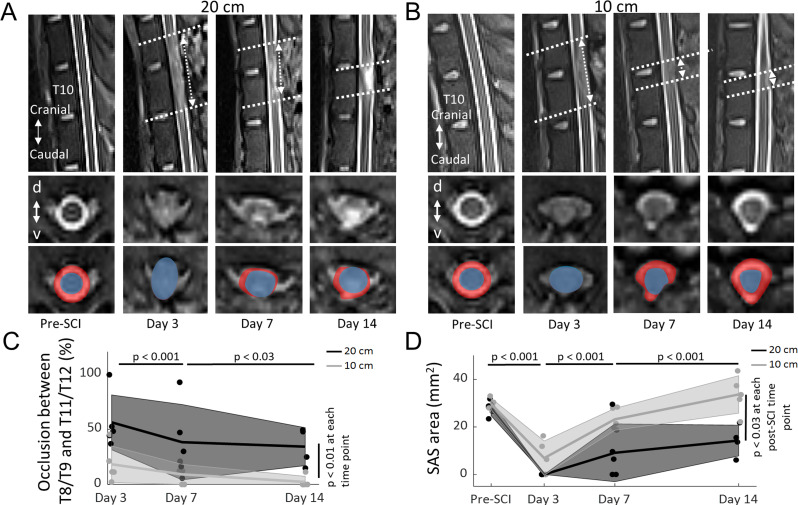
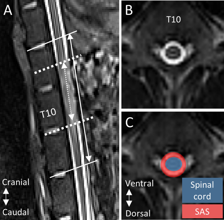
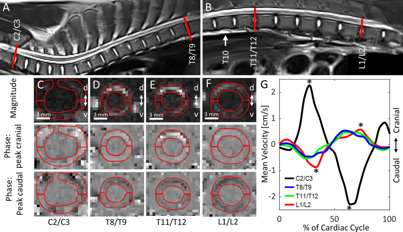
Methods: A thoracic contusion SCI was induced in female domestic pigs (22-29 kg) via a weight drop apparatus (N = 5, 10 cm; N = 5, 20 cm). Magnetic resonance imaging (MRI) was performed pre-SCI and 3, 7 and 14 days post-SCI. SAS occlusion length (cranial-caudal), and injury site SAS area (cross-sectional), were measured on T2-weighted MRI. CSF dynamics, specifically peak cranial/caudal mean velocity (cm/s), and the corresponding time to peak (% of cardiac cycle), were measured on cardiac gated, axial phase-contrast MRI obtained at C2/C3, T8/T9, T11/T12 and L1/L2. Linear-mixed effects models, with a significance level of α = 0.05, were developed to assess the effect of: (1) injury group and time point on SAS occlusion measures; and (2), time point and spinal level, adjusted by injury group, on CSF dynamics.
Results: For both injury groups, SAS occlusion length decreased from 3 to 7 days post-SCI, and 7 to 14 days post-SCI. The cross-sectional SAS area decreased after SCI, and increased to 14 days post-SCI, in both groups. At all spinal levels, peak cranial/caudal mean velocity and the time to peak caudal mean velocity decreased at day 3 post-SCI. From 3 to 14 days post-SCI, peak caudal mean velocity and the time to peak caudal mean velocity increased towards baseline values, at all spinal levels.
Conclusions: Spinal-level specific changes to CSF dynamics, with concurrent changes to SAS occlusion, occurred after SCI in the pig, suggesting that CSF pulsatility and craniospinal compliance were altered in the sub-acute post-traumatic period. These results suggest that PC-MRI derived CSF dynamics may provide a non-invasive method to investigate functional alterations to the spinal intrathecal space following traumatic SCI.
期刊介绍:
"Fluids and Barriers of the CNS" is a scholarly open access journal that specializes in the intricate world of the central nervous system's fluids and barriers, which are pivotal for the health and well-being of the human body. This journal is a peer-reviewed platform that welcomes research manuscripts exploring the full spectrum of CNS fluids and barriers, with a particular focus on their roles in both health and disease.
At the heart of this journal's interest is the cerebrospinal fluid (CSF), a vital fluid that circulates within the brain and spinal cord, playing a multifaceted role in the normal functioning of the brain and in various neurological conditions. The journal delves into the composition, circulation, and absorption of CSF, as well as its relationship with the parenchymal interstitial fluid and the neurovascular unit at the blood-brain barrier (BBB).


 分享
分享
 求助内容:
求助内容: 应助结果提醒方式:
 扫码关注我们
