Ahmed Zakaria, Nessma Sultan, Nesreen Nabil, Mahitabe Elgamily
{"title":"骨髓间充质干细胞衍生的外泌体改善大鼠腮腺唾液腺化疗诱导的损伤。","authors":"Ahmed Zakaria, Nessma Sultan, Nesreen Nabil, Mahitabe Elgamily","doi":"10.1007/s10006-025-01331-9","DOIUrl":null,"url":null,"abstract":"<p><strong>Objective: </strong>A nanometer-sized vesicles originating from bone marrow mesenchymal stem cells (BMMSCs), called exosomes, have been extensively recognized. This study defines the impact of BMMSCs and their derived exosomes on proliferation, apoptosis and oxidative stress (OS) levels of CP-induced parotid salivary gland damage.</p><p><strong>Methods: </strong>BMMSCs were isolated from the tibia of four white albino rats and further characterized by flowcytometric analysis. BMMSCs-derived exosomes were harvested and underwent characterization using transmission electron microscopy (TEM), western blot analysis and BCA assay. Fifty-six healthy white albino male rats weighting from 200 to 250 g were allocated into 4 groups (n = 14); Group I, rats received phosphate buffered saline (PBS), group II, rats were intraperitoneally injected with CP, group III& IV received CP and after 3 days they were intravenously injected with either BMMSCs (group III) or BMMSCs-exosomes (group IV). Histological, and immunohistochemical studies using proliferating cell nuclear antigen (PCNA) were done after 7 and 14 days. The OS was measured using malondialdehyde (MDA) and apoptosis was measured by annexin V-FITC/PI.</p><p><strong>Results: </strong>BMMSCs and exosomes treated groups showed better histological features approximating the normal architecture of the control group. The percentage of PCNA positively stained cells were significantly higher in the exosomes treated group in comparison to all other groups. MDA assay test revealed that the exosomes were able to reduce the OS when compared to the cell-based therapy using BMMSCs. Annexin V revealed that BMMSCs-exosomes significantly reduced the percentage of apoptotic cells compared to other treated groups.</p><p><strong>Conclusions: </strong>BMMSCs-exosomes could improve the CP-induced cytotoxicity in rats' parotid salivary gland.</p>","PeriodicalId":47251,"journal":{"name":"Oral and Maxillofacial Surgery-Heidelberg","volume":"29 1","pages":"39"},"PeriodicalIF":1.8000,"publicationDate":"2025-01-17","publicationTypes":"Journal Article","fieldsOfStudy":null,"isOpenAccess":false,"openAccessPdf":"https://www.ncbi.nlm.nih.gov/pmc/articles/PMC11742274/pdf/","citationCount":"0","resultStr":"{\"title\":\"Exosomes derived from bone marrow mesenchymal stem cells ameliorate chemotherapeutically induced damage in rats' parotid salivary gland.\",\"authors\":\"Ahmed Zakaria, Nessma Sultan, Nesreen Nabil, Mahitabe Elgamily\",\"doi\":\"10.1007/s10006-025-01331-9\",\"DOIUrl\":null,\"url\":null,\"abstract\":\"<p><strong>Objective: </strong>A nanometer-sized vesicles originating from bone marrow mesenchymal stem cells (BMMSCs), called exosomes, have been extensively recognized. This study defines the impact of BMMSCs and their derived exosomes on proliferation, apoptosis and oxidative stress (OS) levels of CP-induced parotid salivary gland damage.</p><p><strong>Methods: </strong>BMMSCs were isolated from the tibia of four white albino rats and further characterized by flowcytometric analysis. BMMSCs-derived exosomes were harvested and underwent characterization using transmission electron microscopy (TEM), western blot analysis and BCA assay. Fifty-six healthy white albino male rats weighting from 200 to 250 g were allocated into 4 groups (n = 14); Group I, rats received phosphate buffered saline (PBS), group II, rats were intraperitoneally injected with CP, group III& IV received CP and after 3 days they were intravenously injected with either BMMSCs (group III) or BMMSCs-exosomes (group IV). Histological, and immunohistochemical studies using proliferating cell nuclear antigen (PCNA) were done after 7 and 14 days. The OS was measured using malondialdehyde (MDA) and apoptosis was measured by annexin V-FITC/PI.</p><p><strong>Results: </strong>BMMSCs and exosomes treated groups showed better histological features approximating the normal architecture of the control group. The percentage of PCNA positively stained cells were significantly higher in the exosomes treated group in comparison to all other groups. MDA assay test revealed that the exosomes were able to reduce the OS when compared to the cell-based therapy using BMMSCs. Annexin V revealed that BMMSCs-exosomes significantly reduced the percentage of apoptotic cells compared to other treated groups.</p><p><strong>Conclusions: </strong>BMMSCs-exosomes could improve the CP-induced cytotoxicity in rats' parotid salivary gland.</p>\",\"PeriodicalId\":47251,\"journal\":{\"name\":\"Oral and Maxillofacial Surgery-Heidelberg\",\"volume\":\"29 1\",\"pages\":\"39\"},\"PeriodicalIF\":1.8000,\"publicationDate\":\"2025-01-17\",\"publicationTypes\":\"Journal Article\",\"fieldsOfStudy\":null,\"isOpenAccess\":false,\"openAccessPdf\":\"https://www.ncbi.nlm.nih.gov/pmc/articles/PMC11742274/pdf/\",\"citationCount\":\"0\",\"resultStr\":null,\"platform\":\"Semanticscholar\",\"paperid\":null,\"PeriodicalName\":\"Oral and Maxillofacial Surgery-Heidelberg\",\"FirstCategoryId\":\"1085\",\"ListUrlMain\":\"https://doi.org/10.1007/s10006-025-01331-9\",\"RegionNum\":0,\"RegionCategory\":null,\"ArticlePicture\":[],\"TitleCN\":null,\"AbstractTextCN\":null,\"PMCID\":null,\"EPubDate\":\"\",\"PubModel\":\"\",\"JCR\":\"Q3\",\"JCRName\":\"DENTISTRY, ORAL SURGERY & MEDICINE\",\"Score\":null,\"Total\":0}","platform":"Semanticscholar","paperid":null,"PeriodicalName":"Oral and Maxillofacial Surgery-Heidelberg","FirstCategoryId":"1085","ListUrlMain":"https://doi.org/10.1007/s10006-025-01331-9","RegionNum":0,"RegionCategory":null,"ArticlePicture":[],"TitleCN":null,"AbstractTextCN":null,"PMCID":null,"EPubDate":"","PubModel":"","JCR":"Q3","JCRName":"DENTISTRY, ORAL SURGERY & MEDICINE","Score":null,"Total":0}
Exosomes derived from bone marrow mesenchymal stem cells ameliorate chemotherapeutically induced damage in rats' parotid salivary gland.
Objective: A nanometer-sized vesicles originating from bone marrow mesenchymal stem cells (BMMSCs), called exosomes, have been extensively recognized. This study defines the impact of BMMSCs and their derived exosomes on proliferation, apoptosis and oxidative stress (OS) levels of CP-induced parotid salivary gland damage.
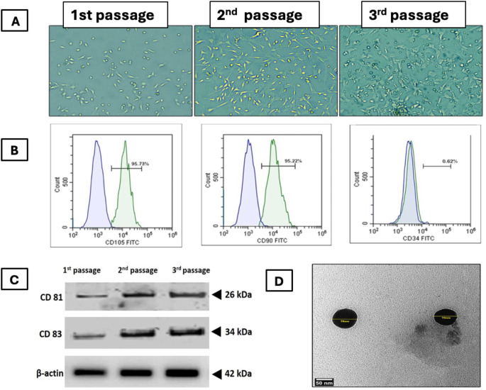
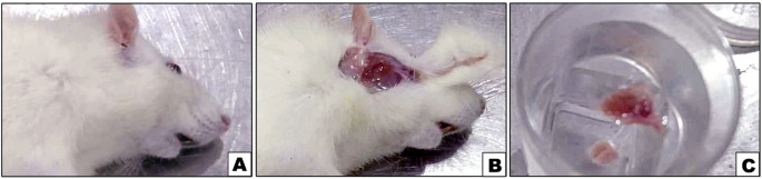
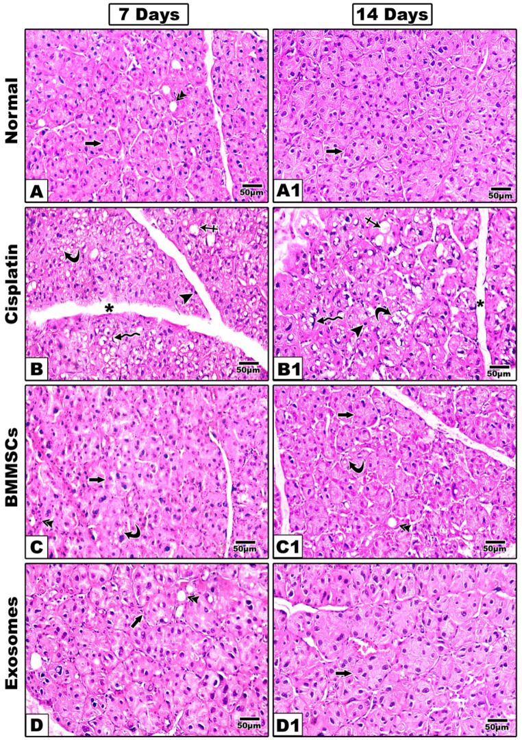
Methods: BMMSCs were isolated from the tibia of four white albino rats and further characterized by flowcytometric analysis. BMMSCs-derived exosomes were harvested and underwent characterization using transmission electron microscopy (TEM), western blot analysis and BCA assay. Fifty-six healthy white albino male rats weighting from 200 to 250 g were allocated into 4 groups (n = 14); Group I, rats received phosphate buffered saline (PBS), group II, rats were intraperitoneally injected with CP, group III& IV received CP and after 3 days they were intravenously injected with either BMMSCs (group III) or BMMSCs-exosomes (group IV). Histological, and immunohistochemical studies using proliferating cell nuclear antigen (PCNA) were done after 7 and 14 days. The OS was measured using malondialdehyde (MDA) and apoptosis was measured by annexin V-FITC/PI.
Results: BMMSCs and exosomes treated groups showed better histological features approximating the normal architecture of the control group. The percentage of PCNA positively stained cells were significantly higher in the exosomes treated group in comparison to all other groups. MDA assay test revealed that the exosomes were able to reduce the OS when compared to the cell-based therapy using BMMSCs. Annexin V revealed that BMMSCs-exosomes significantly reduced the percentage of apoptotic cells compared to other treated groups.
Conclusions: BMMSCs-exosomes could improve the CP-induced cytotoxicity in rats' parotid salivary gland.
期刊介绍:
Oral & Maxillofacial Surgery founded as Mund-, Kiefer- und Gesichtschirurgie is a peer-reviewed online journal. It is designed for clinicians as well as researchers.The quarterly journal offers comprehensive coverage of new techniques, important developments and innovative ideas in oral and maxillofacial surgery and interdisciplinary aspects of cranial, facial and oral diseases and their management. The journal publishes papers of the highest scientific merit and widest possible scope on work in oral and maxillofacial surgery as well as supporting specialties. Practice-oriented articles help improve the methods used in oral and maxillofacial surgery.Every aspect of oral and maxillofacial surgery is fully covered through a range of invited review articles, clinical and research articles, technical notes, abstracts, and case reports. Specific topics are: aesthetic facial surgery, clinical pathology, computer-assisted surgery, congenital and craniofacial deformities, dentoalveolar surgery, head and neck oncology, implant dentistry, oral medicine, orthognathic surgery, reconstructive surgery, skull base surgery, TMJ and trauma.Time-limited reviewing and electronic processing allow to publish articles as fast as possible. Accepted articles are rapidly accessible online.Clinical studies submitted for publication have to include a declaration that they have been approved by an ethical committee according to the World Medical Association Declaration of Helsinki 1964 (last amendment during the 52nd World Medical Association General Assembly, Edinburgh, Scotland, October 2000). Experimental animal studies have to be carried out according to the principles of laboratory animal care (NIH publication No 86-23, revised 1985).


 分享
分享
 求助内容:
求助内容: 应助结果提醒方式:
 扫码关注我们
