{"title":"从三维旋转血管造影虚拟三维重建复杂的先天性心脏解剖结构。","authors":"Ernesto Mejia, Shannon Sweeney, Jenny E Zablah","doi":"10.1186/s41205-024-00247-6","DOIUrl":null,"url":null,"abstract":"<p><strong>Background: </strong>Despite advancements in imaging technologies, including CT scans and MRI, these modalities may still fail to capture intricate details of congenital heart defects accurately. Virtual 3D models have revolutionized the field of pediatric interventional cardiology by providing clinicians with tangible representations of complex anatomical structures. We examined the feasibility and accuracy of utilizing an automated, Artificial Intelligence (AI) driven, cloud-based platform for virtual 3D visualization of complex congenital heart disease obtained from 3D rotational angiography DICOM images.</p><p><strong>Methods: </strong>Five patients selected at random with 3DRA performed in the pediatric cardiac catheterization suite were selected. 3DRA's were performed following published institutional protocols and segmentations performed by primary operators. The 3DRA DICOM images were anonymized as per protocol and exported. Images when then processed by Axial3D Artificial Intelligence (AI) driven cloud-based platform for virtual segmentation. Two separate expert operators were selected to subjectively analyze the segmentations and compare them to the operator reconstructions for anatomic accuracy.</p><p><strong>Results: </strong>Comparing results with local reconstructions by expert operators, five different patient anatomies were analyzed, showcasing Axial3D's ability to produce highly detailed reconstructions with improved visual appeal, including color-coded segments for implanted materials like stents. The reconstructions exhibited superior segmentation of different intrathoracic structures when compared to local models, offering valuable insights for medical professionals and patients.</p><p><strong>Conclusions: </strong>The use of the AI driven, cloud-based platform for 3D visualization of complex congenital heart lesions presents a promising advancement in pediatric interventional cardiology, facilitating enhanced patient care, procedural planning, and educational opportunities for trainees and patients alike.</p>","PeriodicalId":72036,"journal":{"name":"3D printing in medicine","volume":"11 1","pages":"4"},"PeriodicalIF":3.1000,"publicationDate":"2025-01-27","publicationTypes":"Journal Article","fieldsOfStudy":null,"isOpenAccess":false,"openAccessPdf":"https://www.ncbi.nlm.nih.gov/pmc/articles/PMC11770958/pdf/","citationCount":"0","resultStr":"{\"title\":\"Virtual 3D reconstruction of complex congenital cardiac anatomy from 3D rotational angiography.\",\"authors\":\"Ernesto Mejia, Shannon Sweeney, Jenny E Zablah\",\"doi\":\"10.1186/s41205-024-00247-6\",\"DOIUrl\":null,\"url\":null,\"abstract\":\"<p><strong>Background: </strong>Despite advancements in imaging technologies, including CT scans and MRI, these modalities may still fail to capture intricate details of congenital heart defects accurately. Virtual 3D models have revolutionized the field of pediatric interventional cardiology by providing clinicians with tangible representations of complex anatomical structures. We examined the feasibility and accuracy of utilizing an automated, Artificial Intelligence (AI) driven, cloud-based platform for virtual 3D visualization of complex congenital heart disease obtained from 3D rotational angiography DICOM images.</p><p><strong>Methods: </strong>Five patients selected at random with 3DRA performed in the pediatric cardiac catheterization suite were selected. 3DRA's were performed following published institutional protocols and segmentations performed by primary operators. The 3DRA DICOM images were anonymized as per protocol and exported. Images when then processed by Axial3D Artificial Intelligence (AI) driven cloud-based platform for virtual segmentation. Two separate expert operators were selected to subjectively analyze the segmentations and compare them to the operator reconstructions for anatomic accuracy.</p><p><strong>Results: </strong>Comparing results with local reconstructions by expert operators, five different patient anatomies were analyzed, showcasing Axial3D's ability to produce highly detailed reconstructions with improved visual appeal, including color-coded segments for implanted materials like stents. The reconstructions exhibited superior segmentation of different intrathoracic structures when compared to local models, offering valuable insights for medical professionals and patients.</p><p><strong>Conclusions: </strong>The use of the AI driven, cloud-based platform for 3D visualization of complex congenital heart lesions presents a promising advancement in pediatric interventional cardiology, facilitating enhanced patient care, procedural planning, and educational opportunities for trainees and patients alike.</p>\",\"PeriodicalId\":72036,\"journal\":{\"name\":\"3D printing in medicine\",\"volume\":\"11 1\",\"pages\":\"4\"},\"PeriodicalIF\":3.1000,\"publicationDate\":\"2025-01-27\",\"publicationTypes\":\"Journal Article\",\"fieldsOfStudy\":null,\"isOpenAccess\":false,\"openAccessPdf\":\"https://www.ncbi.nlm.nih.gov/pmc/articles/PMC11770958/pdf/\",\"citationCount\":\"0\",\"resultStr\":null,\"platform\":\"Semanticscholar\",\"paperid\":null,\"PeriodicalName\":\"3D printing in medicine\",\"FirstCategoryId\":\"1085\",\"ListUrlMain\":\"https://doi.org/10.1186/s41205-024-00247-6\",\"RegionNum\":0,\"RegionCategory\":null,\"ArticlePicture\":[],\"TitleCN\":null,\"AbstractTextCN\":null,\"PMCID\":null,\"EPubDate\":\"\",\"PubModel\":\"\",\"JCR\":\"Q1\",\"JCRName\":\"RADIOLOGY, NUCLEAR MEDICINE & MEDICAL IMAGING\",\"Score\":null,\"Total\":0}","platform":"Semanticscholar","paperid":null,"PeriodicalName":"3D printing in medicine","FirstCategoryId":"1085","ListUrlMain":"https://doi.org/10.1186/s41205-024-00247-6","RegionNum":0,"RegionCategory":null,"ArticlePicture":[],"TitleCN":null,"AbstractTextCN":null,"PMCID":null,"EPubDate":"","PubModel":"","JCR":"Q1","JCRName":"RADIOLOGY, NUCLEAR MEDICINE & MEDICAL IMAGING","Score":null,"Total":0}
Virtual 3D reconstruction of complex congenital cardiac anatomy from 3D rotational angiography.
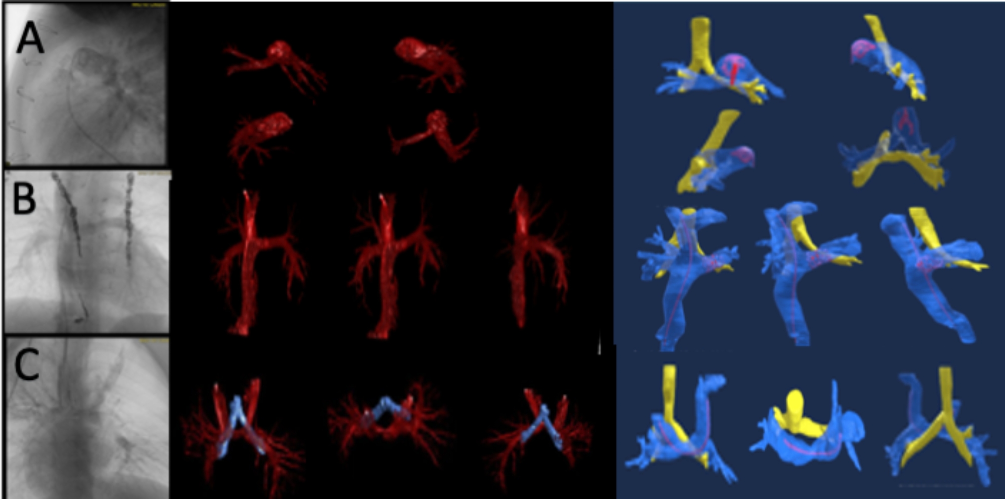
Background: Despite advancements in imaging technologies, including CT scans and MRI, these modalities may still fail to capture intricate details of congenital heart defects accurately. Virtual 3D models have revolutionized the field of pediatric interventional cardiology by providing clinicians with tangible representations of complex anatomical structures. We examined the feasibility and accuracy of utilizing an automated, Artificial Intelligence (AI) driven, cloud-based platform for virtual 3D visualization of complex congenital heart disease obtained from 3D rotational angiography DICOM images.
Methods: Five patients selected at random with 3DRA performed in the pediatric cardiac catheterization suite were selected. 3DRA's were performed following published institutional protocols and segmentations performed by primary operators. The 3DRA DICOM images were anonymized as per protocol and exported. Images when then processed by Axial3D Artificial Intelligence (AI) driven cloud-based platform for virtual segmentation. Two separate expert operators were selected to subjectively analyze the segmentations and compare them to the operator reconstructions for anatomic accuracy.
Results: Comparing results with local reconstructions by expert operators, five different patient anatomies were analyzed, showcasing Axial3D's ability to produce highly detailed reconstructions with improved visual appeal, including color-coded segments for implanted materials like stents. The reconstructions exhibited superior segmentation of different intrathoracic structures when compared to local models, offering valuable insights for medical professionals and patients.
Conclusions: The use of the AI driven, cloud-based platform for 3D visualization of complex congenital heart lesions presents a promising advancement in pediatric interventional cardiology, facilitating enhanced patient care, procedural planning, and educational opportunities for trainees and patients alike.


 分享
分享

 求助内容:
求助内容: 应助结果提醒方式:
 扫码关注我们
