{"title":"评估全身MR图像上骨髓ADC与脂肪抑制方法和脂肪含量的关系。","authors":"Tetsuya Tsujikawa, Akira Makino, Hiroshi Oikawa, Shota Ishida, Tetsuya Mori, Yasushi Kiyono, Hirohiko Kimura, Hidehiko Okazawa","doi":"10.2463/mrms.mp.2020-0129","DOIUrl":null,"url":null,"abstract":"<p><strong>Purpose: </strong>To compare apparent diffusion coefficients (ADCs) of bone marrow on diffusion-weighted imaging (DWI) between two fat-suppression techniques, and to evaluate the association between bone-marrow ADCs and the proton density fat fraction (PDFF).</p><p><strong>Methods: </strong>Seventy-seven patients underwent whole-body DWI with short-inversion time inversion-recovery (STIR) (DWI<sub>STIR</sub>) and/or STIR + selective water-excitation (spectral-spatial RF [SSRF]) (DWI<sub>STIR+SSRF</sub>). ADCs of lumbar vertebrae (L3 and L4) were compared between DWI<sub>STIR</sub> and DWI<sub>STIR+SSRF</sub>, and correlated with the PDFF.</p><p><strong>Results: </strong>Lumbar ADCs obtained by DWI<sub>STIR</sub> and DWI<sub>STIR+SSRF</sub> were significantly correlated (L3: r = 0.90, P < 0.0001, L4: r = 0.90, P < 0.0001). Lumbar ADCs (× 10<sup>-6</sup> mm<sup>2</sup>/s) obtained by DWI<sub>STIR</sub> were significantly lower than those by DWI<sub>STIR+SSRF</sub> (L3: 479 ± 137 and 490 ± 148, P < 0.05, L4: 456 ± 114 and 471 ± 118, P < 0.005). Residual fat signals were more clearly observed on DWI<sub>STIR</sub> than on DWI<sub>STIR+SSRF</sub>. The ADCs of L3 obtained by DWI<sub>STIR</sub> and DWI<sub>STIR+SSRF</sub> exhibited significant positive correlations with the PDFF (r = 0.51, P < 0.0001, and r = 0.45, P < 0.0001, respectively), and the ADCs of L4 obtained by DWI<sub>STIR</sub> and DWI<sub>STIR+SSRF</sub> exhibited significantly positive correlations with the PDFF (r = 0.40, P < 0.0005, and r = 0.40, P < 0.0005, respectively).</p><p><strong>Conclusion: </strong>Irrespective of different fat-suppression methods, lumbar ADCs were positively correlated with the PDFF, being inconsistent with previous studies. Lumbar ADCs obtained by DWI<sub>STIR</sub> were significantly lower than those obtained by DWI<sub>STIR+SSRF,</sub> probably due to residual fat signals on DWI<sub>STIR</sub>. However, this difference (< 4%) did not explain the positive correlation between lumbar ADC and PDFF.</p>","PeriodicalId":18119,"journal":{"name":"Magnetic Resonance in Medical Sciences","volume":"21 3","pages":"407-413"},"PeriodicalIF":3.2000,"publicationDate":"2022-07-01","publicationTypes":"Journal Article","fieldsOfStudy":null,"isOpenAccess":false,"openAccessPdf":"https://ftp.ncbi.nlm.nih.gov/pub/pmc/oa_pdf/a8/15/mrms-21-407.PMC9316130.pdf","citationCount":"1","resultStr":"{\"title\":\"Assessing the ADC of Bone-marrow on Whole-body MR Images in Relation to the Fat-suppression Method and Fat Content.\",\"authors\":\"Tetsuya Tsujikawa, Akira Makino, Hiroshi Oikawa, Shota Ishida, Tetsuya Mori, Yasushi Kiyono, Hirohiko Kimura, Hidehiko Okazawa\",\"doi\":\"10.2463/mrms.mp.2020-0129\",\"DOIUrl\":null,\"url\":null,\"abstract\":\"<p><strong>Purpose: </strong>To compare apparent diffusion coefficients (ADCs) of bone marrow on diffusion-weighted imaging (DWI) between two fat-suppression techniques, and to evaluate the association between bone-marrow ADCs and the proton density fat fraction (PDFF).</p><p><strong>Methods: </strong>Seventy-seven patients underwent whole-body DWI with short-inversion time inversion-recovery (STIR) (DWI<sub>STIR</sub>) and/or STIR + selective water-excitation (spectral-spatial RF [SSRF]) (DWI<sub>STIR+SSRF</sub>). ADCs of lumbar vertebrae (L3 and L4) were compared between DWI<sub>STIR</sub> and DWI<sub>STIR+SSRF</sub>, and correlated with the PDFF.</p><p><strong>Results: </strong>Lumbar ADCs obtained by DWI<sub>STIR</sub> and DWI<sub>STIR+SSRF</sub> were significantly correlated (L3: r = 0.90, P < 0.0001, L4: r = 0.90, P < 0.0001). Lumbar ADCs (× 10<sup>-6</sup> mm<sup>2</sup>/s) obtained by DWI<sub>STIR</sub> were significantly lower than those by DWI<sub>STIR+SSRF</sub> (L3: 479 ± 137 and 490 ± 148, P < 0.05, L4: 456 ± 114 and 471 ± 118, P < 0.005). Residual fat signals were more clearly observed on DWI<sub>STIR</sub> than on DWI<sub>STIR+SSRF</sub>. The ADCs of L3 obtained by DWI<sub>STIR</sub> and DWI<sub>STIR+SSRF</sub> exhibited significant positive correlations with the PDFF (r = 0.51, P < 0.0001, and r = 0.45, P < 0.0001, respectively), and the ADCs of L4 obtained by DWI<sub>STIR</sub> and DWI<sub>STIR+SSRF</sub> exhibited significantly positive correlations with the PDFF (r = 0.40, P < 0.0005, and r = 0.40, P < 0.0005, respectively).</p><p><strong>Conclusion: </strong>Irrespective of different fat-suppression methods, lumbar ADCs were positively correlated with the PDFF, being inconsistent with previous studies. Lumbar ADCs obtained by DWI<sub>STIR</sub> were significantly lower than those obtained by DWI<sub>STIR+SSRF,</sub> probably due to residual fat signals on DWI<sub>STIR</sub>. However, this difference (< 4%) did not explain the positive correlation between lumbar ADC and PDFF.</p>\",\"PeriodicalId\":18119,\"journal\":{\"name\":\"Magnetic Resonance in Medical Sciences\",\"volume\":\"21 3\",\"pages\":\"407-413\"},\"PeriodicalIF\":3.2000,\"publicationDate\":\"2022-07-01\",\"publicationTypes\":\"Journal Article\",\"fieldsOfStudy\":null,\"isOpenAccess\":false,\"openAccessPdf\":\"https://ftp.ncbi.nlm.nih.gov/pub/pmc/oa_pdf/a8/15/mrms-21-407.PMC9316130.pdf\",\"citationCount\":\"1\",\"resultStr\":null,\"platform\":\"Semanticscholar\",\"paperid\":null,\"PeriodicalName\":\"Magnetic Resonance in Medical Sciences\",\"FirstCategoryId\":\"3\",\"ListUrlMain\":\"https://doi.org/10.2463/mrms.mp.2020-0129\",\"RegionNum\":3,\"RegionCategory\":\"医学\",\"ArticlePicture\":[],\"TitleCN\":null,\"AbstractTextCN\":null,\"PMCID\":null,\"EPubDate\":\"2021/2/9 0:00:00\",\"PubModel\":\"Epub\",\"JCR\":\"Q2\",\"JCRName\":\"RADIOLOGY, NUCLEAR MEDICINE & MEDICAL IMAGING\",\"Score\":null,\"Total\":0}","platform":"Semanticscholar","paperid":null,"PeriodicalName":"Magnetic Resonance in Medical Sciences","FirstCategoryId":"3","ListUrlMain":"https://doi.org/10.2463/mrms.mp.2020-0129","RegionNum":3,"RegionCategory":"医学","ArticlePicture":[],"TitleCN":null,"AbstractTextCN":null,"PMCID":null,"EPubDate":"2021/2/9 0:00:00","PubModel":"Epub","JCR":"Q2","JCRName":"RADIOLOGY, NUCLEAR MEDICINE & MEDICAL IMAGING","Score":null,"Total":0}
引用次数: 1
摘要
目的:比较两种脂肪抑制技术在骨髓弥散加权成像(DWI)上的表观扩散系数(adc),探讨骨髓表观扩散系数与质子密度脂肪分数(PDFF)的关系。方法:77例患者采用短反转时间反转恢复(STIR) (DWISTIR)和/或STIR+选择性水激发(光谱-空间RF [SSRF]) (DWISTIR+SSRF)进行全身DWI。比较DWISTIR和DWISTIR+SSRF对腰椎(L3和L4) adc的影响,并与PDFF相关。结果:DWISTIR与DWISTIR+SSRF获得的腰椎adc具有显著相关性(L3: r = 0.90, P < 0.0001; L4: r = 0.90, P < 0.0001)。DWISTIR组腰椎adc (× 10-6 mm2/s)明显低于DWISTIR+SSRF组(L3: 479±137和490±148,P < 0.05, L4: 456±114和471±118,P < 0.005)。与DWISTIR+SSRF相比,DWISTIR组观察到的残余脂肪信号更清晰。DWISTIR和DWISTIR+SSRF获得的L3 adc与PDFF呈显著正相关(r = 0.51, P < 0.0001, r = 0.45, P < 0.0001), DWISTIR和DWISTIR+SSRF获得的L4 adc与PDFF呈显著正相关(r = 0.40, P < 0.0005, r = 0.40, P < 0.0005)。结论:无论采用何种脂肪抑制方法,腰椎adc均与PDFF呈正相关,这与以往的研究不一致。DWISTIR获得的腰椎adc明显低于DWISTIR+SSRF获得的adc,可能是由于DWISTIR上残留的脂肪信号。然而,这一差异(< 4%)并不能解释腰椎ADC和PDFF之间的正相关。
Assessing the ADC of Bone-marrow on Whole-body MR Images in Relation to the Fat-suppression Method and Fat Content.
Purpose: To compare apparent diffusion coefficients (ADCs) of bone marrow on diffusion-weighted imaging (DWI) between two fat-suppression techniques, and to evaluate the association between bone-marrow ADCs and the proton density fat fraction (PDFF).
Methods: Seventy-seven patients underwent whole-body DWI with short-inversion time inversion-recovery (STIR) (DWISTIR) and/or STIR + selective water-excitation (spectral-spatial RF [SSRF]) (DWISTIR+SSRF). ADCs of lumbar vertebrae (L3 and L4) were compared between DWISTIR and DWISTIR+SSRF, and correlated with the PDFF.
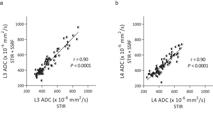
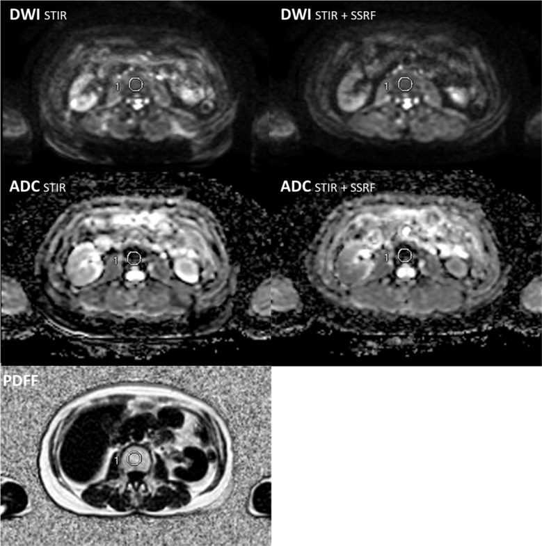
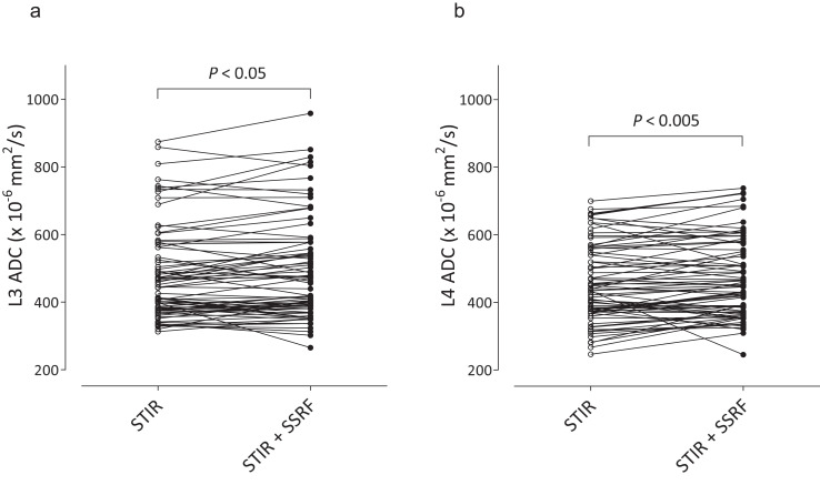
Results: Lumbar ADCs obtained by DWISTIR and DWISTIR+SSRF were significantly correlated (L3: r = 0.90, P < 0.0001, L4: r = 0.90, P < 0.0001). Lumbar ADCs (× 10-6 mm2/s) obtained by DWISTIR were significantly lower than those by DWISTIR+SSRF (L3: 479 ± 137 and 490 ± 148, P < 0.05, L4: 456 ± 114 and 471 ± 118, P < 0.005). Residual fat signals were more clearly observed on DWISTIR than on DWISTIR+SSRF. The ADCs of L3 obtained by DWISTIR and DWISTIR+SSRF exhibited significant positive correlations with the PDFF (r = 0.51, P < 0.0001, and r = 0.45, P < 0.0001, respectively), and the ADCs of L4 obtained by DWISTIR and DWISTIR+SSRF exhibited significantly positive correlations with the PDFF (r = 0.40, P < 0.0005, and r = 0.40, P < 0.0005, respectively).
Conclusion: Irrespective of different fat-suppression methods, lumbar ADCs were positively correlated with the PDFF, being inconsistent with previous studies. Lumbar ADCs obtained by DWISTIR were significantly lower than those obtained by DWISTIR+SSRF, probably due to residual fat signals on DWISTIR. However, this difference (< 4%) did not explain the positive correlation between lumbar ADC and PDFF.
期刊介绍:
Magnetic Resonance in Medical Sciences (MRMS or Magn
Reson Med Sci) is an international journal pursuing the
publication of original articles contributing to the progress
of magnetic resonance in the field of biomedical sciences
including technical developments and clinical applications.
MRMS is an official journal of the Japanese Society for
Magnetic Resonance in Medicine (JSMRM).


 分享
分享
 求助内容:
求助内容: 应助结果提醒方式:
 扫码关注我们
