{"title":"改进接触x射线显微摄影法测量牙硬组织矿物质密度。","authors":"B D Schmuck, C M Carey","doi":"10.6028/jres.115.006","DOIUrl":null,"url":null,"abstract":"<p><p>Contact X-ray microradiography is the current gold standard for measuring mineral densities of partially demineralized tooth specimens. The X-ray sensitive film specified in the last J Res NIST publication on the subject is no longer commercially available. OBJECTIVES: Develop a new microradiographic method by identifying a commercially available film with greater than 3000 lines per millimeter resolution, which is sensitive to X rays, and develop correct film processing for X-ray microradiographic application. METHODS: A holographic film was identified as a potential replacement film. Proper exposure was determined utilizing a thick nickel plate to create test-strips. Film development was bracketed around manufacturer suggestions. Film linearity was determined with aluminum step-wedges. Microradiographs of 100 µm thick tooth sections, before and after acidic challenges, were a final test for film. Magnified images were captured with a digital microscope camera with 0.305 micrometers per pixel resolution. RESULTS: The appropriate film exposure was 30 minutes at 80 kV(p) and 3 mA with a development time of 2 minutes. Step-wedge experiments show the system to be linear in terms of pixel intensities with respect to x-ray attenuation for normalized pixel intensity values that are 10% to 90% of full scale (r(2) = 0.997) which encompasses the full exposure region of tooth tissue. Enamel sections were analyzed and show distinctive differences between erosion and demineralization. The image capture device resolution of 0.305 micrometers per pixel limits the system resolution. CONCLUSION: Use of the identified holographic film when combined with the described processing modifications has resulted in an improved X-ray microradiographic method for the measurement of mineral density of dental hard tissues. The method described can be further improved by using a higher resolution digitization system. The method is appropriate for quantitatively measuring changes in mineral density and erosion.</p>","PeriodicalId":17039,"journal":{"name":"Journal of Research of the National Institute of Standards and Technology","volume":"115 2","pages":"75-83"},"PeriodicalIF":1.5000,"publicationDate":"2010-04-01","publicationTypes":"Journal Article","fieldsOfStudy":null,"isOpenAccess":false,"openAccessPdf":"https://www.ncbi.nlm.nih.gov/pmc/articles/PMC3086209/pdf/","citationCount":"0","resultStr":"{\"title\":\"Improved Contact X-Ray Microradiographic Method to Measure Mineral Density of Hard Dental Tissues.\",\"authors\":\"B D Schmuck, C M Carey\",\"doi\":\"10.6028/jres.115.006\",\"DOIUrl\":null,\"url\":null,\"abstract\":\"<p><p>Contact X-ray microradiography is the current gold standard for measuring mineral densities of partially demineralized tooth specimens. The X-ray sensitive film specified in the last J Res NIST publication on the subject is no longer commercially available. OBJECTIVES: Develop a new microradiographic method by identifying a commercially available film with greater than 3000 lines per millimeter resolution, which is sensitive to X rays, and develop correct film processing for X-ray microradiographic application. METHODS: A holographic film was identified as a potential replacement film. Proper exposure was determined utilizing a thick nickel plate to create test-strips. Film development was bracketed around manufacturer suggestions. Film linearity was determined with aluminum step-wedges. Microradiographs of 100 µm thick tooth sections, before and after acidic challenges, were a final test for film. Magnified images were captured with a digital microscope camera with 0.305 micrometers per pixel resolution. RESULTS: The appropriate film exposure was 30 minutes at 80 kV(p) and 3 mA with a development time of 2 minutes. Step-wedge experiments show the system to be linear in terms of pixel intensities with respect to x-ray attenuation for normalized pixel intensity values that are 10% to 90% of full scale (r(2) = 0.997) which encompasses the full exposure region of tooth tissue. Enamel sections were analyzed and show distinctive differences between erosion and demineralization. The image capture device resolution of 0.305 micrometers per pixel limits the system resolution. CONCLUSION: Use of the identified holographic film when combined with the described processing modifications has resulted in an improved X-ray microradiographic method for the measurement of mineral density of dental hard tissues. The method described can be further improved by using a higher resolution digitization system. The method is appropriate for quantitatively measuring changes in mineral density and erosion.</p>\",\"PeriodicalId\":17039,\"journal\":{\"name\":\"Journal of Research of the National Institute of Standards and Technology\",\"volume\":\"115 2\",\"pages\":\"75-83\"},\"PeriodicalIF\":1.5000,\"publicationDate\":\"2010-04-01\",\"publicationTypes\":\"Journal Article\",\"fieldsOfStudy\":null,\"isOpenAccess\":false,\"openAccessPdf\":\"https://www.ncbi.nlm.nih.gov/pmc/articles/PMC3086209/pdf/\",\"citationCount\":\"0\",\"resultStr\":null,\"platform\":\"Semanticscholar\",\"paperid\":null,\"PeriodicalName\":\"Journal of Research of the National Institute of Standards and Technology\",\"FirstCategoryId\":\"5\",\"ListUrlMain\":\"https://doi.org/10.6028/jres.115.006\",\"RegionNum\":4,\"RegionCategory\":\"工程技术\",\"ArticlePicture\":[],\"TitleCN\":null,\"AbstractTextCN\":null,\"PMCID\":null,\"EPubDate\":\"2010/3/1 0:00:00\",\"PubModel\":\"Print\",\"JCR\":\"\",\"JCRName\":\"\",\"Score\":null,\"Total\":0}","platform":"Semanticscholar","paperid":null,"PeriodicalName":"Journal of Research of the National Institute of Standards and Technology","FirstCategoryId":"5","ListUrlMain":"https://doi.org/10.6028/jres.115.006","RegionNum":4,"RegionCategory":"工程技术","ArticlePicture":[],"TitleCN":null,"AbstractTextCN":null,"PMCID":null,"EPubDate":"2010/3/1 0:00:00","PubModel":"Print","JCR":"","JCRName":"","Score":null,"Total":0}
Improved Contact X-Ray Microradiographic Method to Measure Mineral Density of Hard Dental Tissues.
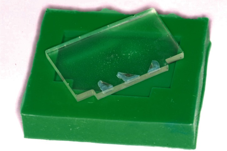
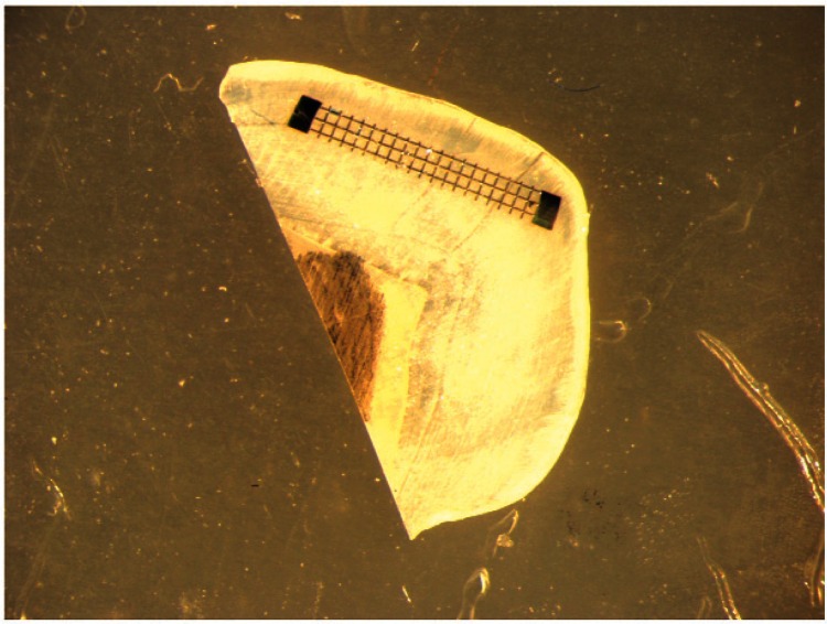
Contact X-ray microradiography is the current gold standard for measuring mineral densities of partially demineralized tooth specimens. The X-ray sensitive film specified in the last J Res NIST publication on the subject is no longer commercially available. OBJECTIVES: Develop a new microradiographic method by identifying a commercially available film with greater than 3000 lines per millimeter resolution, which is sensitive to X rays, and develop correct film processing for X-ray microradiographic application. METHODS: A holographic film was identified as a potential replacement film. Proper exposure was determined utilizing a thick nickel plate to create test-strips. Film development was bracketed around manufacturer suggestions. Film linearity was determined with aluminum step-wedges. Microradiographs of 100 µm thick tooth sections, before and after acidic challenges, were a final test for film. Magnified images were captured with a digital microscope camera with 0.305 micrometers per pixel resolution. RESULTS: The appropriate film exposure was 30 minutes at 80 kV(p) and 3 mA with a development time of 2 minutes. Step-wedge experiments show the system to be linear in terms of pixel intensities with respect to x-ray attenuation for normalized pixel intensity values that are 10% to 90% of full scale (r(2) = 0.997) which encompasses the full exposure region of tooth tissue. Enamel sections were analyzed and show distinctive differences between erosion and demineralization. The image capture device resolution of 0.305 micrometers per pixel limits the system resolution. CONCLUSION: Use of the identified holographic film when combined with the described processing modifications has resulted in an improved X-ray microradiographic method for the measurement of mineral density of dental hard tissues. The method described can be further improved by using a higher resolution digitization system. The method is appropriate for quantitatively measuring changes in mineral density and erosion.
期刊介绍:
The Journal of Research of the National Institute of Standards and Technology is the flagship publication of the National Institute of Standards and Technology. It has been published under various titles and forms since 1904, with its roots as Scientific Papers issued as the Bulletin of the Bureau of Standards.
In 1928, the Scientific Papers were combined with Technologic Papers, which reported results of investigations of material and methods of testing. This new publication was titled the Bureau of Standards Journal of Research.
The Journal of Research of NIST reports NIST research and development in metrology and related fields of physical science, engineering, applied mathematics, statistics, biotechnology, information technology.


 分享
分享
 求助内容:
求助内容: 应助结果提醒方式:
 扫码关注我们
