Achim Fieß, Marilena Brandt, Eva Mildenberger, Michael Siegfried Urschitz, Felix Mathias Wagner, Stephanie Desiree Grabitz, Esther Maria Hoffmann, Norbert Pfeiffer, Alexander Konrad Schuster
{"title":"足月出生时胎龄小的成年人乳头周围视网膜神经纤维层比对照组薄。","authors":"Achim Fieß, Marilena Brandt, Eva Mildenberger, Michael Siegfried Urschitz, Felix Mathias Wagner, Stephanie Desiree Grabitz, Esther Maria Hoffmann, Norbert Pfeiffer, Alexander Konrad Schuster","doi":"10.2147/EB.S383231","DOIUrl":null,"url":null,"abstract":"<p><strong>Purpose: </strong>Prenatal growth restriction is associated with impaired neurodevelopment in childhood. This study investigated the effects of being born small for gestational age (SGA) on peripapillary retinal nerve fiber layer (pRNFL) thickness in adults born at term.</p><p><strong>Methods: </strong>A retrospective cohort study was conducted with a prospective ophthalmologic examination of participants born at full-term (gestational age ≥37 weeks) between 1969 and 2002. All participants were examined with spectral-domain optical coherence tomography and grouped according to their birth weight in correlation to gestational age as former moderate (birth weight (BW) percentile 3rd to <10th) and severe SGA (<3rd percentile), normal (10th-90th percentile, AGA), and moderately (>90th to 97th percentile) and severely (>97th percentile) large for gestational age (LGA) adults (18 to 52 years).</p><p><strong>Results: </strong>Overall, 547 eyes of 285 individuals (age 29.9±9.4 years, 151 females) born at term were included. Multivariable regression analyses revealed a strong association between a lower global pRNFL thickness in the severe SGA (B=-8.99 [95%-CI: -12.68; -5.30] µm; p<0.001) and in the moderate SGA groups (B=-6.40 [95%-CI: -10.29; -2.50] µm; p=0.001) compared to the reference AGA group.</p><p><strong>Conclusion: </strong>Our results indicate that restricted fetal growth affects neurologic tissue development of the optic nerve head, particularly in individuals born severely SGA at term. This indicates that fetal growth restriction may exert disturbances in the development of neurologic tissue, which persists in adulthood.</p>","PeriodicalId":51844,"journal":{"name":"Eye and Brain","volume":null,"pages":null},"PeriodicalIF":3.1000,"publicationDate":"2022-11-25","publicationTypes":"Journal Article","fieldsOfStudy":null,"isOpenAccess":false,"openAccessPdf":"https://ftp.ncbi.nlm.nih.gov/pub/pmc/oa_pdf/3c/df/eb-14-127.PMC9709856.pdf","citationCount":"2","resultStr":"{\"title\":\"Adults Born Small for Gestational Age at Term Have Thinner Peripapillary Retinal Nerve Fiber Layers Than Controls.\",\"authors\":\"Achim Fieß, Marilena Brandt, Eva Mildenberger, Michael Siegfried Urschitz, Felix Mathias Wagner, Stephanie Desiree Grabitz, Esther Maria Hoffmann, Norbert Pfeiffer, Alexander Konrad Schuster\",\"doi\":\"10.2147/EB.S383231\",\"DOIUrl\":null,\"url\":null,\"abstract\":\"<p><strong>Purpose: </strong>Prenatal growth restriction is associated with impaired neurodevelopment in childhood. This study investigated the effects of being born small for gestational age (SGA) on peripapillary retinal nerve fiber layer (pRNFL) thickness in adults born at term.</p><p><strong>Methods: </strong>A retrospective cohort study was conducted with a prospective ophthalmologic examination of participants born at full-term (gestational age ≥37 weeks) between 1969 and 2002. All participants were examined with spectral-domain optical coherence tomography and grouped according to their birth weight in correlation to gestational age as former moderate (birth weight (BW) percentile 3rd to <10th) and severe SGA (<3rd percentile), normal (10th-90th percentile, AGA), and moderately (>90th to 97th percentile) and severely (>97th percentile) large for gestational age (LGA) adults (18 to 52 years).</p><p><strong>Results: </strong>Overall, 547 eyes of 285 individuals (age 29.9±9.4 years, 151 females) born at term were included. Multivariable regression analyses revealed a strong association between a lower global pRNFL thickness in the severe SGA (B=-8.99 [95%-CI: -12.68; -5.30] µm; p<0.001) and in the moderate SGA groups (B=-6.40 [95%-CI: -10.29; -2.50] µm; p=0.001) compared to the reference AGA group.</p><p><strong>Conclusion: </strong>Our results indicate that restricted fetal growth affects neurologic tissue development of the optic nerve head, particularly in individuals born severely SGA at term. This indicates that fetal growth restriction may exert disturbances in the development of neurologic tissue, which persists in adulthood.</p>\",\"PeriodicalId\":51844,\"journal\":{\"name\":\"Eye and Brain\",\"volume\":null,\"pages\":null},\"PeriodicalIF\":3.1000,\"publicationDate\":\"2022-11-25\",\"publicationTypes\":\"Journal Article\",\"fieldsOfStudy\":null,\"isOpenAccess\":false,\"openAccessPdf\":\"https://ftp.ncbi.nlm.nih.gov/pub/pmc/oa_pdf/3c/df/eb-14-127.PMC9709856.pdf\",\"citationCount\":\"2\",\"resultStr\":null,\"platform\":\"Semanticscholar\",\"paperid\":null,\"PeriodicalName\":\"Eye and Brain\",\"FirstCategoryId\":\"1085\",\"ListUrlMain\":\"https://doi.org/10.2147/EB.S383231\",\"RegionNum\":0,\"RegionCategory\":null,\"ArticlePicture\":[],\"TitleCN\":null,\"AbstractTextCN\":null,\"PMCID\":null,\"EPubDate\":\"2022/1/1 0:00:00\",\"PubModel\":\"eCollection\",\"JCR\":\"Q1\",\"JCRName\":\"OPHTHALMOLOGY\",\"Score\":null,\"Total\":0}","platform":"Semanticscholar","paperid":null,"PeriodicalName":"Eye and Brain","FirstCategoryId":"1085","ListUrlMain":"https://doi.org/10.2147/EB.S383231","RegionNum":0,"RegionCategory":null,"ArticlePicture":[],"TitleCN":null,"AbstractTextCN":null,"PMCID":null,"EPubDate":"2022/1/1 0:00:00","PubModel":"eCollection","JCR":"Q1","JCRName":"OPHTHALMOLOGY","Score":null,"Total":0}
Adults Born Small for Gestational Age at Term Have Thinner Peripapillary Retinal Nerve Fiber Layers Than Controls.
Purpose: Prenatal growth restriction is associated with impaired neurodevelopment in childhood. This study investigated the effects of being born small for gestational age (SGA) on peripapillary retinal nerve fiber layer (pRNFL) thickness in adults born at term.
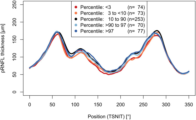
Methods: A retrospective cohort study was conducted with a prospective ophthalmologic examination of participants born at full-term (gestational age ≥37 weeks) between 1969 and 2002. All participants were examined with spectral-domain optical coherence tomography and grouped according to their birth weight in correlation to gestational age as former moderate (birth weight (BW) percentile 3rd to <10th) and severe SGA (<3rd percentile), normal (10th-90th percentile, AGA), and moderately (>90th to 97th percentile) and severely (>97th percentile) large for gestational age (LGA) adults (18 to 52 years).
Results: Overall, 547 eyes of 285 individuals (age 29.9±9.4 years, 151 females) born at term were included. Multivariable regression analyses revealed a strong association between a lower global pRNFL thickness in the severe SGA (B=-8.99 [95%-CI: -12.68; -5.30] µm; p<0.001) and in the moderate SGA groups (B=-6.40 [95%-CI: -10.29; -2.50] µm; p=0.001) compared to the reference AGA group.
Conclusion: Our results indicate that restricted fetal growth affects neurologic tissue development of the optic nerve head, particularly in individuals born severely SGA at term. This indicates that fetal growth restriction may exert disturbances in the development of neurologic tissue, which persists in adulthood.
期刊介绍:
Eye and Brain is an international, peer-reviewed, open access journal focusing on basic research, clinical findings, and expert reviews in the field of visual science and neuro-ophthalmology. The journal’s unique focus is the link between two well-known visual centres, the eye and the brain, with an emphasis on the importance of such connections. All aspects of clinical and especially basic research on the visual system are addressed within the journal as well as significant future directions in vision research and therapeutic measures. This unique journal focuses on neurological aspects of vision – both physiological and pathological. The scope of the journal spans from the cornea to the associational visual cortex and all the visual centers in between. Topics range from basic biological mechanisms to therapeutic treatment, from simple organisms to humans, and utilizing techniques from molecular biology to behavior. The journal especially welcomes primary research articles or review papers that make the connection between the eye and the brain. Specific areas covered in the journal include: Physiology and pathophysiology of visual centers, Eye movement disorders and strabismus, Cellular, biochemical, and molecular features of the visual system, Structural and functional organization of the eye and of the visual cortex, Metabolic demands of the visual system, Diseases and disorders with neuro-ophthalmic manifestations, Clinical and experimental neuro-ophthalmology and visual system pathologies, Epidemiological studies.


 分享
分享
 求助内容:
求助内容: 应助结果提醒方式:
 扫码关注我们
