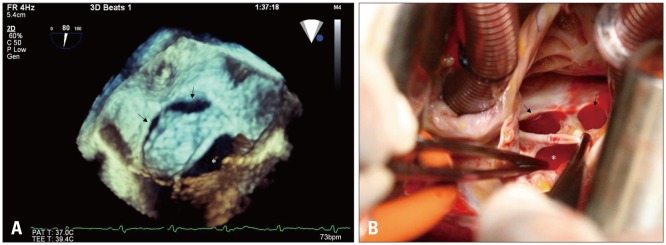
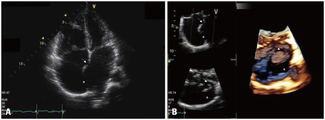
{"title":"经食道三维超声心动图证实马蹄状房间隔缺损。","authors":"Jin-Sun Park, Joon-Han Shin","doi":"10.4250/jcu.2017.25.4.138","DOIUrl":null,"url":null,"abstract":"A 51-year-old female with exertional dyspnea was admitted. Transthoracic echocardiography (TTE) revealed 2 defects at interatrial septum with left to right shunt (Fig. 1A). En face display of the interatrial septum by three-dimensional (3D) TTE demonstrated two ovoid atrial septal defects (ASDs). Two ovoid defects and interatrial septum formed like horseshoe (Fig. 1B). Transesophageal echocardiography (TEE) revealed total three oval shaped ASDs, which were well-visualized in one view, using 3D volume rendering of the interatrial septum from real-time 3D data (Fig. 2A). This finding was consistent with an intraoperative image (Fig. 2B). 3D TEE has a distinct advantage over two-dimensional (2D) echocardiography in case of complex ASD, especially when 2 or more defects are present. 3D echocardiography can provide pISSN 1975-4612 / eISSN 2005-9655 Copyright © 2017 Korean Society of Echocardiography www.kse-jcu.org https://doi.org/10.4250/jcu.2017.25.4.138","PeriodicalId":88913,"journal":{"name":"Journal of cardiovascular ultrasound","volume":"25 4","pages":"138-139"},"PeriodicalIF":0.0000,"publicationDate":"2017-12-01","publicationTypes":"Journal Article","fieldsOfStudy":null,"isOpenAccess":false,"openAccessPdf":"https://sci-hub-pdf.com/10.4250/jcu.2017.25.4.138","citationCount":"0","resultStr":"{\"title\":\"Horseshoe-like Shaped Atrial Septal Defects Confirmed on Three-Dimensional Transesophageal Echocardiography.\",\"authors\":\"Jin-Sun Park, Joon-Han Shin\",\"doi\":\"10.4250/jcu.2017.25.4.138\",\"DOIUrl\":null,\"url\":null,\"abstract\":\"A 51-year-old female with exertional dyspnea was admitted. Transthoracic echocardiography (TTE) revealed 2 defects at interatrial septum with left to right shunt (Fig. 1A). En face display of the interatrial septum by three-dimensional (3D) TTE demonstrated two ovoid atrial septal defects (ASDs). Two ovoid defects and interatrial septum formed like horseshoe (Fig. 1B). Transesophageal echocardiography (TEE) revealed total three oval shaped ASDs, which were well-visualized in one view, using 3D volume rendering of the interatrial septum from real-time 3D data (Fig. 2A). This finding was consistent with an intraoperative image (Fig. 2B). 3D TEE has a distinct advantage over two-dimensional (2D) echocardiography in case of complex ASD, especially when 2 or more defects are present. 3D echocardiography can provide pISSN 1975-4612 / eISSN 2005-9655 Copyright © 2017 Korean Society of Echocardiography www.kse-jcu.org https://doi.org/10.4250/jcu.2017.25.4.138\",\"PeriodicalId\":88913,\"journal\":{\"name\":\"Journal of cardiovascular ultrasound\",\"volume\":\"25 4\",\"pages\":\"138-139\"},\"PeriodicalIF\":0.0000,\"publicationDate\":\"2017-12-01\",\"publicationTypes\":\"Journal Article\",\"fieldsOfStudy\":null,\"isOpenAccess\":false,\"openAccessPdf\":\"https://sci-hub-pdf.com/10.4250/jcu.2017.25.4.138\",\"citationCount\":\"0\",\"resultStr\":null,\"platform\":\"Semanticscholar\",\"paperid\":null,\"PeriodicalName\":\"Journal of cardiovascular ultrasound\",\"FirstCategoryId\":\"1085\",\"ListUrlMain\":\"https://doi.org/10.4250/jcu.2017.25.4.138\",\"RegionNum\":0,\"RegionCategory\":null,\"ArticlePicture\":[],\"TitleCN\":null,\"AbstractTextCN\":null,\"PMCID\":null,\"EPubDate\":\"2017/12/29 0:00:00\",\"PubModel\":\"Epub\",\"JCR\":\"\",\"JCRName\":\"\",\"Score\":null,\"Total\":0}","platform":"Semanticscholar","paperid":null,"PeriodicalName":"Journal of cardiovascular ultrasound","FirstCategoryId":"1085","ListUrlMain":"https://doi.org/10.4250/jcu.2017.25.4.138","RegionNum":0,"RegionCategory":null,"ArticlePicture":[],"TitleCN":null,"AbstractTextCN":null,"PMCID":null,"EPubDate":"2017/12/29 0:00:00","PubModel":"Epub","JCR":"","JCRName":"","Score":null,"Total":0}
引用次数: 0


