{"title":"外侧膝状核的连接组入路。","authors":"Josh L Morgan","doi":"10.1017/S0952523817000116","DOIUrl":null,"url":null,"abstract":"<p><p>Although the core functions and structure of the lateral geniculate nucleus (LGN) are well understood, this core is surrounded by questions about the integration of feedforward and feedback connections, interactions between different channels of information, and how activity dependent development restructures synaptic networks. Our understanding of the organization of the mouse LGN is particularly limited given how important it has become as a model system. Advances in circuit scale electron microscopy (cellular connectomics) have made it possible to reconstruct the synaptic connectivity of hundreds of neurons within in a circuit the size of the mouse LGN. These circuit reconstructions can reveal cell type-to-cell type canonical wiring diagrams as well as the higher order wiring motifs that are only visible in reconstructions of intact networks. Connectomic analysis of the LGN therefore not only can answer longstanding questions about the organization of the visual thalamus but also presents unique opportunities for investigating fundamental properties of mammalian circuit formation.</p>","PeriodicalId":23556,"journal":{"name":"Visual Neuroscience","volume":"34 ","pages":"E014"},"PeriodicalIF":2.3000,"publicationDate":"2017-01-01","publicationTypes":"Journal Article","fieldsOfStudy":null,"isOpenAccess":false,"openAccessPdf":"https://sci-hub-pdf.com/10.1017/S0952523817000116","citationCount":"2","resultStr":"{\"title\":\"A connectomic approach to the lateral geniculate nucleus.\",\"authors\":\"Josh L Morgan\",\"doi\":\"10.1017/S0952523817000116\",\"DOIUrl\":null,\"url\":null,\"abstract\":\"<p><p>Although the core functions and structure of the lateral geniculate nucleus (LGN) are well understood, this core is surrounded by questions about the integration of feedforward and feedback connections, interactions between different channels of information, and how activity dependent development restructures synaptic networks. Our understanding of the organization of the mouse LGN is particularly limited given how important it has become as a model system. Advances in circuit scale electron microscopy (cellular connectomics) have made it possible to reconstruct the synaptic connectivity of hundreds of neurons within in a circuit the size of the mouse LGN. These circuit reconstructions can reveal cell type-to-cell type canonical wiring diagrams as well as the higher order wiring motifs that are only visible in reconstructions of intact networks. Connectomic analysis of the LGN therefore not only can answer longstanding questions about the organization of the visual thalamus but also presents unique opportunities for investigating fundamental properties of mammalian circuit formation.</p>\",\"PeriodicalId\":23556,\"journal\":{\"name\":\"Visual Neuroscience\",\"volume\":\"34 \",\"pages\":\"E014\"},\"PeriodicalIF\":2.3000,\"publicationDate\":\"2017-01-01\",\"publicationTypes\":\"Journal Article\",\"fieldsOfStudy\":null,\"isOpenAccess\":false,\"openAccessPdf\":\"https://sci-hub-pdf.com/10.1017/S0952523817000116\",\"citationCount\":\"2\",\"resultStr\":null,\"platform\":\"Semanticscholar\",\"paperid\":null,\"PeriodicalName\":\"Visual Neuroscience\",\"FirstCategoryId\":\"3\",\"ListUrlMain\":\"https://doi.org/10.1017/S0952523817000116\",\"RegionNum\":4,\"RegionCategory\":\"医学\",\"ArticlePicture\":[],\"TitleCN\":null,\"AbstractTextCN\":null,\"PMCID\":null,\"EPubDate\":\"\",\"PubModel\":\"\",\"JCR\":\"Q4\",\"JCRName\":\"NEUROSCIENCES\",\"Score\":null,\"Total\":0}","platform":"Semanticscholar","paperid":null,"PeriodicalName":"Visual Neuroscience","FirstCategoryId":"3","ListUrlMain":"https://doi.org/10.1017/S0952523817000116","RegionNum":4,"RegionCategory":"医学","ArticlePicture":[],"TitleCN":null,"AbstractTextCN":null,"PMCID":null,"EPubDate":"","PubModel":"","JCR":"Q4","JCRName":"NEUROSCIENCES","Score":null,"Total":0}
A connectomic approach to the lateral geniculate nucleus.
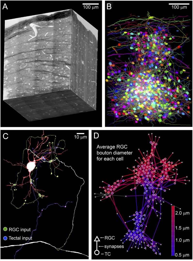
Although the core functions and structure of the lateral geniculate nucleus (LGN) are well understood, this core is surrounded by questions about the integration of feedforward and feedback connections, interactions between different channels of information, and how activity dependent development restructures synaptic networks. Our understanding of the organization of the mouse LGN is particularly limited given how important it has become as a model system. Advances in circuit scale electron microscopy (cellular connectomics) have made it possible to reconstruct the synaptic connectivity of hundreds of neurons within in a circuit the size of the mouse LGN. These circuit reconstructions can reveal cell type-to-cell type canonical wiring diagrams as well as the higher order wiring motifs that are only visible in reconstructions of intact networks. Connectomic analysis of the LGN therefore not only can answer longstanding questions about the organization of the visual thalamus but also presents unique opportunities for investigating fundamental properties of mammalian circuit formation.
期刊介绍:
Visual Neuroscience is an international journal devoted to the publication of experimental and theoretical research on biological mechanisms of vision. A major goal of publication is to bring together in one journal a broad range of studies that reflect the diversity and originality of all aspects of neuroscience research relating to the visual system. Contributions may address molecular, cellular or systems-level processes in either vertebrate or invertebrate species. The journal publishes work based on a wide range of technical approaches, including molecular genetics, anatomy, physiology, psychophysics and imaging, and utilizing comparative, developmental, theoretical or computational approaches to understand the biology of vision and visuo-motor control. The journal also publishes research seeking to understand disorders of the visual system and strategies for restoring vision. Studies based exclusively on clinical, psychophysiological or behavioral data are welcomed, provided that they address questions concerning neural mechanisms of vision or provide insight into visual dysfunction.


 分享
分享
 求助内容:
求助内容: 应助结果提醒方式:
 扫码关注我们
