{"title":"模仿肝细胞癌的肝脏原发性副神经节瘤。","authors":"Chia-Shuen Lin, Yung-Hsiang Hsu","doi":"10.4103/tcmj.tcmj_230_18","DOIUrl":null,"url":null,"abstract":"<p><p>Paragangliomas occurring primarily in the liver parenchyma are extremely rare. The radiological features mimic hepatocellular carcinoma. Herein, we describe a case with a presurgical diagnosis of hepatocellular carcinoma, which was pathologically confirmed as hepatic paraganglioma after surgery. A 41-year-old woman visited our hospital for evaluation of a hepatic mass detected incidentally in a health examination 7 years previously. Triple-phase computed tomography of the liver showed a homogeneous and strongly arterial enhancing mass, followed by portovenous washout at the posterior segment of the right lobe. In view of the enhancement pattern and highly vascular nature, the hepatocellular carcinoma was included in the differential diagnosis. Anatomical resection of segment VII was carried out. Grossly, the mass was encapsulated and soft. Microscopically, it had a typical \"Zellballen pattern.\" Immunohistochemical staining for CD56 was positive, and sustentacular cells at the periphery of the tumor cell nests were also demonstrated by S-100 protein. Therefore, the diagnosis of primary paraganglioma of the liver was confirmed. In conclusion, hepatic paraganglioma should be considered in the differential diagnosis in cases of hepatic masses displaying \"early enhancement, early washout.\"</p>","PeriodicalId":72593,"journal":{"name":"Ci ji yi xue za zhi = Tzu-chi medical journal","volume":"31 4","pages":"286-288"},"PeriodicalIF":0.0000,"publicationDate":"2019-09-16","publicationTypes":"Journal Article","fieldsOfStudy":null,"isOpenAccess":false,"openAccessPdf":"https://ftp.ncbi.nlm.nih.gov/pub/pmc/oa_pdf/6a/f7/TCMJ-31-286.PMC6905243.pdf","citationCount":"0","resultStr":"{\"title\":\"A primary paraganglioma of the liver mimicking hepatocellular carcinoma.\",\"authors\":\"Chia-Shuen Lin, Yung-Hsiang Hsu\",\"doi\":\"10.4103/tcmj.tcmj_230_18\",\"DOIUrl\":null,\"url\":null,\"abstract\":\"<p><p>Paragangliomas occurring primarily in the liver parenchyma are extremely rare. The radiological features mimic hepatocellular carcinoma. Herein, we describe a case with a presurgical diagnosis of hepatocellular carcinoma, which was pathologically confirmed as hepatic paraganglioma after surgery. A 41-year-old woman visited our hospital for evaluation of a hepatic mass detected incidentally in a health examination 7 years previously. Triple-phase computed tomography of the liver showed a homogeneous and strongly arterial enhancing mass, followed by portovenous washout at the posterior segment of the right lobe. In view of the enhancement pattern and highly vascular nature, the hepatocellular carcinoma was included in the differential diagnosis. Anatomical resection of segment VII was carried out. Grossly, the mass was encapsulated and soft. Microscopically, it had a typical \\\"Zellballen pattern.\\\" Immunohistochemical staining for CD56 was positive, and sustentacular cells at the periphery of the tumor cell nests were also demonstrated by S-100 protein. Therefore, the diagnosis of primary paraganglioma of the liver was confirmed. In conclusion, hepatic paraganglioma should be considered in the differential diagnosis in cases of hepatic masses displaying \\\"early enhancement, early washout.\\\"</p>\",\"PeriodicalId\":72593,\"journal\":{\"name\":\"Ci ji yi xue za zhi = Tzu-chi medical journal\",\"volume\":\"31 4\",\"pages\":\"286-288\"},\"PeriodicalIF\":0.0000,\"publicationDate\":\"2019-09-16\",\"publicationTypes\":\"Journal Article\",\"fieldsOfStudy\":null,\"isOpenAccess\":false,\"openAccessPdf\":\"https://ftp.ncbi.nlm.nih.gov/pub/pmc/oa_pdf/6a/f7/TCMJ-31-286.PMC6905243.pdf\",\"citationCount\":\"0\",\"resultStr\":null,\"platform\":\"Semanticscholar\",\"paperid\":null,\"PeriodicalName\":\"Ci ji yi xue za zhi = Tzu-chi medical journal\",\"FirstCategoryId\":\"1085\",\"ListUrlMain\":\"https://doi.org/10.4103/tcmj.tcmj_230_18\",\"RegionNum\":0,\"RegionCategory\":null,\"ArticlePicture\":[],\"TitleCN\":null,\"AbstractTextCN\":null,\"PMCID\":null,\"EPubDate\":\"2019/10/1 0:00:00\",\"PubModel\":\"eCollection\",\"JCR\":\"\",\"JCRName\":\"\",\"Score\":null,\"Total\":0}","platform":"Semanticscholar","paperid":null,"PeriodicalName":"Ci ji yi xue za zhi = Tzu-chi medical journal","FirstCategoryId":"1085","ListUrlMain":"https://doi.org/10.4103/tcmj.tcmj_230_18","RegionNum":0,"RegionCategory":null,"ArticlePicture":[],"TitleCN":null,"AbstractTextCN":null,"PMCID":null,"EPubDate":"2019/10/1 0:00:00","PubModel":"eCollection","JCR":"","JCRName":"","Score":null,"Total":0}
A primary paraganglioma of the liver mimicking hepatocellular carcinoma.
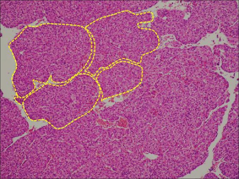
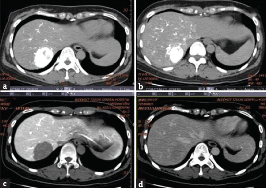
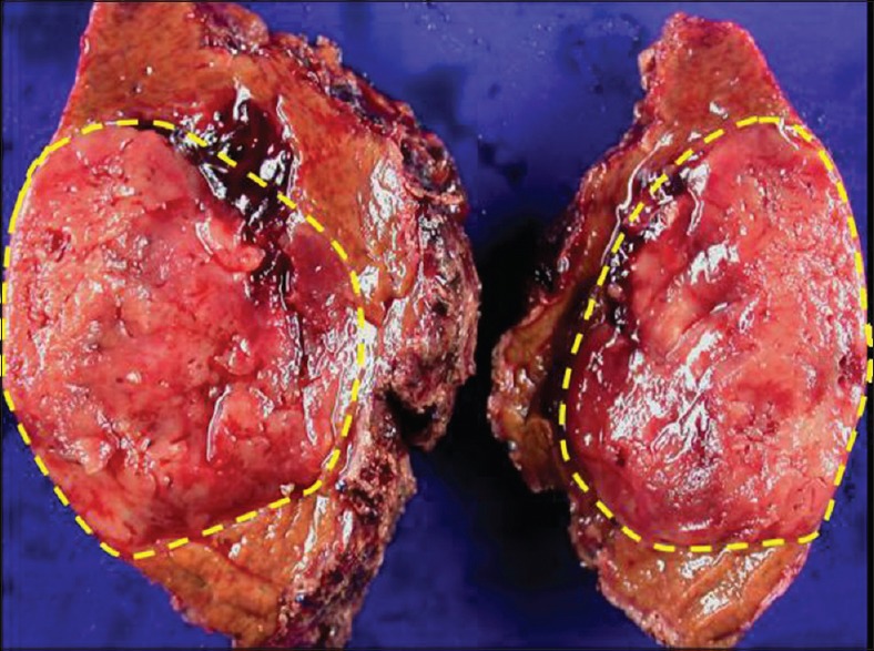
Paragangliomas occurring primarily in the liver parenchyma are extremely rare. The radiological features mimic hepatocellular carcinoma. Herein, we describe a case with a presurgical diagnosis of hepatocellular carcinoma, which was pathologically confirmed as hepatic paraganglioma after surgery. A 41-year-old woman visited our hospital for evaluation of a hepatic mass detected incidentally in a health examination 7 years previously. Triple-phase computed tomography of the liver showed a homogeneous and strongly arterial enhancing mass, followed by portovenous washout at the posterior segment of the right lobe. In view of the enhancement pattern and highly vascular nature, the hepatocellular carcinoma was included in the differential diagnosis. Anatomical resection of segment VII was carried out. Grossly, the mass was encapsulated and soft. Microscopically, it had a typical "Zellballen pattern." Immunohistochemical staining for CD56 was positive, and sustentacular cells at the periphery of the tumor cell nests were also demonstrated by S-100 protein. Therefore, the diagnosis of primary paraganglioma of the liver was confirmed. In conclusion, hepatic paraganglioma should be considered in the differential diagnosis in cases of hepatic masses displaying "early enhancement, early washout."


 分享
分享

 求助内容:
求助内容: 应助结果提醒方式:
 扫码关注我们
