Philip Procter, Michael Pujari-Palmer, Gry Hulsart-Billström, David Wenner, Gerard Insley, Sune Larsson, Håkan Engqvist
{"title":"评价骨软骨组织粘接剂的生物力学试验模型。","authors":"Philip Procter, Michael Pujari-Palmer, Gry Hulsart-Billström, David Wenner, Gerard Insley, Sune Larsson, Håkan Engqvist","doi":"10.1186/s42490-019-0011-2","DOIUrl":null,"url":null,"abstract":"<p><strong>Background: </strong>Currently there are no standard models with which to evaluate the biomechanical performance of calcified tissue adhesives, in vivo<i>.</i> We present, herein, a pre-clinical murine distal femoral bone model for evaluating tissue adhesives intended for use in both osseous and osteochondral tissue reconstruction.</p><p><strong>Results: </strong>Cylindrical cores (diameter (Ø) 2 mm (mm) × 2 mm depth), containing both cancellous and cortical bone, were fractured out from the distal femur and then reattached using one of two tissue adhesives. The adhesiveness of fibrin glue (Tisseel<sup>tm</sup>), and a novel, biocompatible, calcium phosphate-based tissue adhesive (OsStic<sup>tm</sup>) were evaluated by pullout testing, in which glued cores were extracted and the peak force at failure recorded. The results show that Tisseel weakly bonded the metaphyseal bone cores, while OsStic produced > 30-fold higher mean peak forces at failure (7.64 Newtons (N) vs. 0.21 N). The failure modes were consistently disparate, with Tisseel failing gradually, while OsStic failed abruptly, as would be expected with a calcium-based material. Imaging of the bone/adhesive interface with microcomputed tomography revealed that, for OsStic, failure occurred more often within cancellous bone (75% of tested samples) rather than at the adhesive interface.</p><p><strong>Conclusions: </strong>Despite the challenges associated with biomechanical testing in small rodent models the preclinical ex-vivo test model presented herein is both sensitive and accurate. It enabled differences in tissue adhesive strength to be quantified even for very small osseous fragments (<Ø4mm). Importantly, this model can easily be scaled to larger animals and adapted to fracture fragment fixation in human bone. The present model is also compatible with other long-term in vivo evaluation methods (i.e. in vivo imaging, histological analysis, etc.).</p>","PeriodicalId":72425,"journal":{"name":"BMC biomedical engineering","volume":"1 ","pages":"11"},"PeriodicalIF":0.0000,"publicationDate":"2019-05-07","publicationTypes":"Journal Article","fieldsOfStudy":null,"isOpenAccess":false,"openAccessPdf":"https://sci-hub-pdf.com/10.1186/s42490-019-0011-2","citationCount":"17","resultStr":"{\"title\":\"A biomechanical test model for evaluating osseous and osteochondral tissue adhesives.\",\"authors\":\"Philip Procter, Michael Pujari-Palmer, Gry Hulsart-Billström, David Wenner, Gerard Insley, Sune Larsson, Håkan Engqvist\",\"doi\":\"10.1186/s42490-019-0011-2\",\"DOIUrl\":null,\"url\":null,\"abstract\":\"<p><strong>Background: </strong>Currently there are no standard models with which to evaluate the biomechanical performance of calcified tissue adhesives, in vivo<i>.</i> We present, herein, a pre-clinical murine distal femoral bone model for evaluating tissue adhesives intended for use in both osseous and osteochondral tissue reconstruction.</p><p><strong>Results: </strong>Cylindrical cores (diameter (Ø) 2 mm (mm) × 2 mm depth), containing both cancellous and cortical bone, were fractured out from the distal femur and then reattached using one of two tissue adhesives. The adhesiveness of fibrin glue (Tisseel<sup>tm</sup>), and a novel, biocompatible, calcium phosphate-based tissue adhesive (OsStic<sup>tm</sup>) were evaluated by pullout testing, in which glued cores were extracted and the peak force at failure recorded. The results show that Tisseel weakly bonded the metaphyseal bone cores, while OsStic produced > 30-fold higher mean peak forces at failure (7.64 Newtons (N) vs. 0.21 N). The failure modes were consistently disparate, with Tisseel failing gradually, while OsStic failed abruptly, as would be expected with a calcium-based material. Imaging of the bone/adhesive interface with microcomputed tomography revealed that, for OsStic, failure occurred more often within cancellous bone (75% of tested samples) rather than at the adhesive interface.</p><p><strong>Conclusions: </strong>Despite the challenges associated with biomechanical testing in small rodent models the preclinical ex-vivo test model presented herein is both sensitive and accurate. It enabled differences in tissue adhesive strength to be quantified even for very small osseous fragments (<Ø4mm). Importantly, this model can easily be scaled to larger animals and adapted to fracture fragment fixation in human bone. The present model is also compatible with other long-term in vivo evaluation methods (i.e. in vivo imaging, histological analysis, etc.).</p>\",\"PeriodicalId\":72425,\"journal\":{\"name\":\"BMC biomedical engineering\",\"volume\":\"1 \",\"pages\":\"11\"},\"PeriodicalIF\":0.0000,\"publicationDate\":\"2019-05-07\",\"publicationTypes\":\"Journal Article\",\"fieldsOfStudy\":null,\"isOpenAccess\":false,\"openAccessPdf\":\"https://sci-hub-pdf.com/10.1186/s42490-019-0011-2\",\"citationCount\":\"17\",\"resultStr\":null,\"platform\":\"Semanticscholar\",\"paperid\":null,\"PeriodicalName\":\"BMC biomedical engineering\",\"FirstCategoryId\":\"1085\",\"ListUrlMain\":\"https://doi.org/10.1186/s42490-019-0011-2\",\"RegionNum\":0,\"RegionCategory\":null,\"ArticlePicture\":[],\"TitleCN\":null,\"AbstractTextCN\":null,\"PMCID\":null,\"EPubDate\":\"2019/1/1 0:00:00\",\"PubModel\":\"eCollection\",\"JCR\":\"\",\"JCRName\":\"\",\"Score\":null,\"Total\":0}","platform":"Semanticscholar","paperid":null,"PeriodicalName":"BMC biomedical engineering","FirstCategoryId":"1085","ListUrlMain":"https://doi.org/10.1186/s42490-019-0011-2","RegionNum":0,"RegionCategory":null,"ArticlePicture":[],"TitleCN":null,"AbstractTextCN":null,"PMCID":null,"EPubDate":"2019/1/1 0:00:00","PubModel":"eCollection","JCR":"","JCRName":"","Score":null,"Total":0}
A biomechanical test model for evaluating osseous and osteochondral tissue adhesives.
Background: Currently there are no standard models with which to evaluate the biomechanical performance of calcified tissue adhesives, in vivo. We present, herein, a pre-clinical murine distal femoral bone model for evaluating tissue adhesives intended for use in both osseous and osteochondral tissue reconstruction.
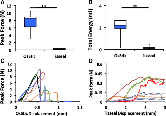
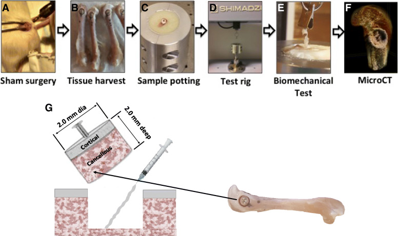
Results: Cylindrical cores (diameter (Ø) 2 mm (mm) × 2 mm depth), containing both cancellous and cortical bone, were fractured out from the distal femur and then reattached using one of two tissue adhesives. The adhesiveness of fibrin glue (Tisseeltm), and a novel, biocompatible, calcium phosphate-based tissue adhesive (OsStictm) were evaluated by pullout testing, in which glued cores were extracted and the peak force at failure recorded. The results show that Tisseel weakly bonded the metaphyseal bone cores, while OsStic produced > 30-fold higher mean peak forces at failure (7.64 Newtons (N) vs. 0.21 N). The failure modes were consistently disparate, with Tisseel failing gradually, while OsStic failed abruptly, as would be expected with a calcium-based material. Imaging of the bone/adhesive interface with microcomputed tomography revealed that, for OsStic, failure occurred more often within cancellous bone (75% of tested samples) rather than at the adhesive interface.
Conclusions: Despite the challenges associated with biomechanical testing in small rodent models the preclinical ex-vivo test model presented herein is both sensitive and accurate. It enabled differences in tissue adhesive strength to be quantified even for very small osseous fragments (<Ø4mm). Importantly, this model can easily be scaled to larger animals and adapted to fracture fragment fixation in human bone. The present model is also compatible with other long-term in vivo evaluation methods (i.e. in vivo imaging, histological analysis, etc.).


 分享
分享

 求助内容:
求助内容: 应助结果提醒方式:
 扫码关注我们
