{"title":"基于深度卷积神经网络的组织病理学图像自动细胞核分割方法。","authors":"Hwejin Jung, Bilal Lodhi, Jaewoo Kang","doi":"10.1186/s42490-019-0026-8","DOIUrl":null,"url":null,"abstract":"<p><strong>Background: </strong>Since nuclei segmentation in histopathology images can provide key information for identifying the presence or stage of a disease, the images need to be assessed carefully. However, color variation in histopathology images, and various structures of nuclei are two major obstacles in accurately segmenting and analyzing histopathology images. Several machine learning methods heavily rely on hand-crafted features which have limitations due to manual thresholding.</p><p><strong>Results: </strong>To obtain robust results, deep learning based methods have been proposed. Deep convolutional neural networks (DCNN) used for automatically extracting features from raw image data have been proven to achieve great performance. Inspired by such achievements, we propose a nuclei segmentation method based on DCNNs. To normalize the color of histopathology images, we use a deep convolutional Gaussian mixture color normalization model which is able to cluster pixels while considering the structures of nuclei. To segment nuclei, we use Mask R-CNN which achieves state-of-the-art object segmentation performance in the field of computer vision. In addition, we perform multiple inference as a post-processing step to boost segmentation performance. We evaluate our segmentation method on two different datasets. The first dataset consists of histopathology images of various organ while the other consists histopathology images of the same organ. Performance of our segmentation method is measured in various experimental setups at the object-level and the pixel-level. In addition, we compare the performance of our method with that of existing state-of-the-art methods. The experimental results show that our nuclei segmentation method outperforms the existing methods.</p><p><strong>Conclusions: </strong>We propose a nuclei segmentation method based on DCNNs for histopathology images. The proposed method which uses Mask R-CNN with color normalization and multiple inference post-processing provides robust nuclei segmentation results. Our method also can facilitate downstream nuclei morphological analyses as it provides high-quality features extracted from histopathology images.</p>","PeriodicalId":72425,"journal":{"name":"BMC biomedical engineering","volume":"1 ","pages":"24"},"PeriodicalIF":0.0000,"publicationDate":"2019-10-17","publicationTypes":"Journal Article","fieldsOfStudy":null,"isOpenAccess":false,"openAccessPdf":"https://www.ncbi.nlm.nih.gov/pmc/articles/PMC7422516/pdf/","citationCount":"0","resultStr":"{\"title\":\"An automatic nuclei segmentation method based on deep convolutional neural networks for histopathology images.\",\"authors\":\"Hwejin Jung, Bilal Lodhi, Jaewoo Kang\",\"doi\":\"10.1186/s42490-019-0026-8\",\"DOIUrl\":null,\"url\":null,\"abstract\":\"<p><strong>Background: </strong>Since nuclei segmentation in histopathology images can provide key information for identifying the presence or stage of a disease, the images need to be assessed carefully. However, color variation in histopathology images, and various structures of nuclei are two major obstacles in accurately segmenting and analyzing histopathology images. Several machine learning methods heavily rely on hand-crafted features which have limitations due to manual thresholding.</p><p><strong>Results: </strong>To obtain robust results, deep learning based methods have been proposed. Deep convolutional neural networks (DCNN) used for automatically extracting features from raw image data have been proven to achieve great performance. Inspired by such achievements, we propose a nuclei segmentation method based on DCNNs. To normalize the color of histopathology images, we use a deep convolutional Gaussian mixture color normalization model which is able to cluster pixels while considering the structures of nuclei. To segment nuclei, we use Mask R-CNN which achieves state-of-the-art object segmentation performance in the field of computer vision. In addition, we perform multiple inference as a post-processing step to boost segmentation performance. We evaluate our segmentation method on two different datasets. The first dataset consists of histopathology images of various organ while the other consists histopathology images of the same organ. Performance of our segmentation method is measured in various experimental setups at the object-level and the pixel-level. In addition, we compare the performance of our method with that of existing state-of-the-art methods. The experimental results show that our nuclei segmentation method outperforms the existing methods.</p><p><strong>Conclusions: </strong>We propose a nuclei segmentation method based on DCNNs for histopathology images. The proposed method which uses Mask R-CNN with color normalization and multiple inference post-processing provides robust nuclei segmentation results. Our method also can facilitate downstream nuclei morphological analyses as it provides high-quality features extracted from histopathology images.</p>\",\"PeriodicalId\":72425,\"journal\":{\"name\":\"BMC biomedical engineering\",\"volume\":\"1 \",\"pages\":\"24\"},\"PeriodicalIF\":0.0000,\"publicationDate\":\"2019-10-17\",\"publicationTypes\":\"Journal Article\",\"fieldsOfStudy\":null,\"isOpenAccess\":false,\"openAccessPdf\":\"https://www.ncbi.nlm.nih.gov/pmc/articles/PMC7422516/pdf/\",\"citationCount\":\"0\",\"resultStr\":null,\"platform\":\"Semanticscholar\",\"paperid\":null,\"PeriodicalName\":\"BMC biomedical engineering\",\"FirstCategoryId\":\"1085\",\"ListUrlMain\":\"https://doi.org/10.1186/s42490-019-0026-8\",\"RegionNum\":0,\"RegionCategory\":null,\"ArticlePicture\":[],\"TitleCN\":null,\"AbstractTextCN\":null,\"PMCID\":null,\"EPubDate\":\"2019/1/1 0:00:00\",\"PubModel\":\"eCollection\",\"JCR\":\"\",\"JCRName\":\"\",\"Score\":null,\"Total\":0}","platform":"Semanticscholar","paperid":null,"PeriodicalName":"BMC biomedical engineering","FirstCategoryId":"1085","ListUrlMain":"https://doi.org/10.1186/s42490-019-0026-8","RegionNum":0,"RegionCategory":null,"ArticlePicture":[],"TitleCN":null,"AbstractTextCN":null,"PMCID":null,"EPubDate":"2019/1/1 0:00:00","PubModel":"eCollection","JCR":"","JCRName":"","Score":null,"Total":0}
An automatic nuclei segmentation method based on deep convolutional neural networks for histopathology images.
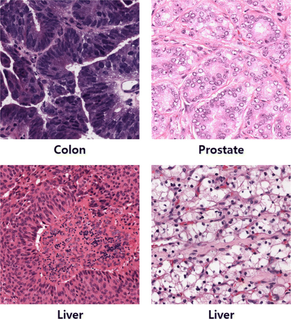
Background: Since nuclei segmentation in histopathology images can provide key information for identifying the presence or stage of a disease, the images need to be assessed carefully. However, color variation in histopathology images, and various structures of nuclei are two major obstacles in accurately segmenting and analyzing histopathology images. Several machine learning methods heavily rely on hand-crafted features which have limitations due to manual thresholding.
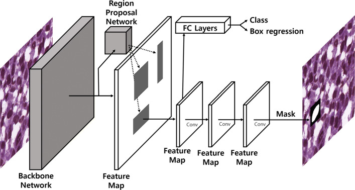
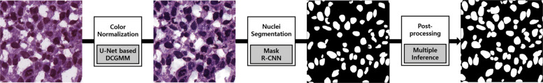
Results: To obtain robust results, deep learning based methods have been proposed. Deep convolutional neural networks (DCNN) used for automatically extracting features from raw image data have been proven to achieve great performance. Inspired by such achievements, we propose a nuclei segmentation method based on DCNNs. To normalize the color of histopathology images, we use a deep convolutional Gaussian mixture color normalization model which is able to cluster pixels while considering the structures of nuclei. To segment nuclei, we use Mask R-CNN which achieves state-of-the-art object segmentation performance in the field of computer vision. In addition, we perform multiple inference as a post-processing step to boost segmentation performance. We evaluate our segmentation method on two different datasets. The first dataset consists of histopathology images of various organ while the other consists histopathology images of the same organ. Performance of our segmentation method is measured in various experimental setups at the object-level and the pixel-level. In addition, we compare the performance of our method with that of existing state-of-the-art methods. The experimental results show that our nuclei segmentation method outperforms the existing methods.
Conclusions: We propose a nuclei segmentation method based on DCNNs for histopathology images. The proposed method which uses Mask R-CNN with color normalization and multiple inference post-processing provides robust nuclei segmentation results. Our method also can facilitate downstream nuclei morphological analyses as it provides high-quality features extracted from histopathology images.


 分享
分享

 求助内容:
求助内容: 应助结果提醒方式:
 扫码关注我们
