{"title":"利用FIB-SEM研究细胞器动力学在光镜活体成像和电子显微镜三维结构之间的相关性","authors":"Keisuke Ohta;Shingo Hirashima;Yoshihiro Miyazono;Akinobu Togo;Kei-ichiro Nakamura","doi":"10.1093/jmicro/dfaa071","DOIUrl":null,"url":null,"abstract":"Correlative light and electron microscopy (CLEM) methods combined with live imaging can be applied to understand the dynamics of organelles. Although recent advances in cell biology and light microscopy have helped in visualizing the details of organelle activities, observing their ultrastructure or organization of surrounding microenvironments is a challenging task. Therefore, CLEM, which allows us to observe the same area as an optical microscope with an electron microscope, has become a key technique in cell biology. Unfortunately, most CLEM methods have technical drawbacks, and many researchers face difficulties in applying CLEM methods. Here, we propose a live three-dimensional CLEM method, combined with a three-dimensional reconstruction technique using focused ion beam scanning electron microscopy tomography, as a solution to such technical barriers. We review our method, the associated technical limitations and the options considered to perform live CLEM.","PeriodicalId":18515,"journal":{"name":"Microscopy","volume":"70 1","pages":"161-170"},"PeriodicalIF":1.8000,"publicationDate":"2020-11-01","publicationTypes":"Journal Article","fieldsOfStudy":null,"isOpenAccess":false,"openAccessPdf":"https://sci-hub-pdf.com/10.1093/jmicro/dfaa071","citationCount":"7","resultStr":"{\"title\":\"Correlation of organelle dynamics between light microscopic live imaging and electron microscopic 3D architecture using FIB-SEM\",\"authors\":\"Keisuke Ohta;Shingo Hirashima;Yoshihiro Miyazono;Akinobu Togo;Kei-ichiro Nakamura\",\"doi\":\"10.1093/jmicro/dfaa071\",\"DOIUrl\":null,\"url\":null,\"abstract\":\"Correlative light and electron microscopy (CLEM) methods combined with live imaging can be applied to understand the dynamics of organelles. Although recent advances in cell biology and light microscopy have helped in visualizing the details of organelle activities, observing their ultrastructure or organization of surrounding microenvironments is a challenging task. Therefore, CLEM, which allows us to observe the same area as an optical microscope with an electron microscope, has become a key technique in cell biology. Unfortunately, most CLEM methods have technical drawbacks, and many researchers face difficulties in applying CLEM methods. Here, we propose a live three-dimensional CLEM method, combined with a three-dimensional reconstruction technique using focused ion beam scanning electron microscopy tomography, as a solution to such technical barriers. We review our method, the associated technical limitations and the options considered to perform live CLEM.\",\"PeriodicalId\":18515,\"journal\":{\"name\":\"Microscopy\",\"volume\":\"70 1\",\"pages\":\"161-170\"},\"PeriodicalIF\":1.8000,\"publicationDate\":\"2020-11-01\",\"publicationTypes\":\"Journal Article\",\"fieldsOfStudy\":null,\"isOpenAccess\":false,\"openAccessPdf\":\"https://sci-hub-pdf.com/10.1093/jmicro/dfaa071\",\"citationCount\":\"7\",\"resultStr\":null,\"platform\":\"Semanticscholar\",\"paperid\":null,\"PeriodicalName\":\"Microscopy\",\"FirstCategoryId\":\"5\",\"ListUrlMain\":\"https://ieeexplore.ieee.org/document/9433138/\",\"RegionNum\":4,\"RegionCategory\":\"工程技术\",\"ArticlePicture\":[],\"TitleCN\":null,\"AbstractTextCN\":null,\"PMCID\":null,\"EPubDate\":\"\",\"PubModel\":\"\",\"JCR\":\"\",\"JCRName\":\"\",\"Score\":null,\"Total\":0}","platform":"Semanticscholar","paperid":null,"PeriodicalName":"Microscopy","FirstCategoryId":"5","ListUrlMain":"https://ieeexplore.ieee.org/document/9433138/","RegionNum":4,"RegionCategory":"工程技术","ArticlePicture":[],"TitleCN":null,"AbstractTextCN":null,"PMCID":null,"EPubDate":"","PubModel":"","JCR":"","JCRName":"","Score":null,"Total":0}
Correlation of organelle dynamics between light microscopic live imaging and electron microscopic 3D architecture using FIB-SEM
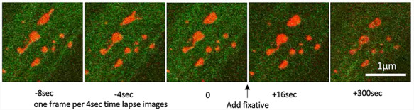
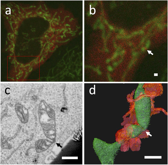
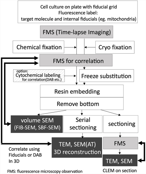
Correlative light and electron microscopy (CLEM) methods combined with live imaging can be applied to understand the dynamics of organelles. Although recent advances in cell biology and light microscopy have helped in visualizing the details of organelle activities, observing their ultrastructure or organization of surrounding microenvironments is a challenging task. Therefore, CLEM, which allows us to observe the same area as an optical microscope with an electron microscope, has become a key technique in cell biology. Unfortunately, most CLEM methods have technical drawbacks, and many researchers face difficulties in applying CLEM methods. Here, we propose a live three-dimensional CLEM method, combined with a three-dimensional reconstruction technique using focused ion beam scanning electron microscopy tomography, as a solution to such technical barriers. We review our method, the associated technical limitations and the options considered to perform live CLEM.
期刊介绍:
Microscopy, previously Journal of Electron Microscopy, promotes research combined with any type of microscopy techniques, applied in life and material sciences. Microscopy is the official journal of the Japanese Society of Microscopy.


 分享
分享
 求助内容:
求助内容: 应助结果提醒方式:
 扫码关注我们
