{"title":"DCE-MRI与t1wi鉴别附件恶性肿块的诊断价值。","authors":"Satoshi Otani, Aki Kido, Yuki Himoto, Akihiko Sakata, Tomoaki Otani, Ryo Kuwahara, Yusaku Moribata, Naoko Nishio, Ryo Yajima, Kyoko Nakao, Yasuhisa Kurata, Sachiko Minamiguchi, Masaki Mandai, Yuji Nakamoto","doi":"10.2463/mrms.mp.2021-0003","DOIUrl":null,"url":null,"abstract":"<p><strong>Purpose: </strong>To compare the diagnostic performance of dynamic contrast-enhanced-MR (DCE-MR) and delayed contrast-enhanced (CE)-MRI added to unenhanced MRI, including diffusion weighted image (DWI) for differentiating malignant adnexal tumors, conducting a retrospective blinded image interpretation study.</p><p><strong>Methods: </strong>Data of 80 patients suspected of having adnexal tumors by ultrasonography between April 2008 and August 2018 were used for the study. All patients had undergone preoperative MRI and surgical resection at our institution. Four radiologists (two specialized in gynecological radiology and two non-specialized) were enrolled for blinded review of the MR images. A 3-point scale was used: 0 = benign, 1 = indeterminate, and 2 = malignant. Three imaging sets were reviewed: Set A, unenhanced MRI including DWI; Set B, Set A and delayed CE-T1WI; and Set C, Set A and DCE-MRI. Imaging criteria for benign and malignant tumors were given in earlier reports. The diagnostic performance of the three imaging sets of the four readers was calculated. Their areas under the curve (AUCs) were compared using the DeLong method.</p><p><strong>Results: </strong>Accuracies of Set B were 81%-88%. Those of Set C were 81%-85%. The AUCs of Set B were 0.83 and 0.89. Those of Set C were 0.81-0.86. For two readers, Set A showed lower accuracy and AUC than Set B/Set C (less than 0.80), although those were equivalent in other readers. No significant difference in AUCs was found among the three sequence sets. Intrareader agreement was moderate to almost perfect in Sets A and B, and substantial to almost perfect in Set C.</p><p><strong>Conclusion: </strong>DCE-MR showed no superiority for differentiating malignant adnexal tumors from benign tumors compared to delayed CE-T1WI with conventional MR and DWI.</p>","PeriodicalId":18119,"journal":{"name":"Magnetic Resonance in Medical Sciences","volume":"21 4","pages":"599-607"},"PeriodicalIF":3.2000,"publicationDate":"2022-10-01","publicationTypes":"Journal Article","fieldsOfStudy":null,"isOpenAccess":false,"openAccessPdf":"https://ftp.ncbi.nlm.nih.gov/pub/pmc/oa_pdf/a1/8d/mrms-21-599.PMC9618924.pdf","citationCount":"3","resultStr":"{\"title\":\"Diagnostic Value of DCE-MRI for Differentiating Malignant Adnexal Masses Compared with Contrast-enhanced-T1WI.\",\"authors\":\"Satoshi Otani, Aki Kido, Yuki Himoto, Akihiko Sakata, Tomoaki Otani, Ryo Kuwahara, Yusaku Moribata, Naoko Nishio, Ryo Yajima, Kyoko Nakao, Yasuhisa Kurata, Sachiko Minamiguchi, Masaki Mandai, Yuji Nakamoto\",\"doi\":\"10.2463/mrms.mp.2021-0003\",\"DOIUrl\":null,\"url\":null,\"abstract\":\"<p><strong>Purpose: </strong>To compare the diagnostic performance of dynamic contrast-enhanced-MR (DCE-MR) and delayed contrast-enhanced (CE)-MRI added to unenhanced MRI, including diffusion weighted image (DWI) for differentiating malignant adnexal tumors, conducting a retrospective blinded image interpretation study.</p><p><strong>Methods: </strong>Data of 80 patients suspected of having adnexal tumors by ultrasonography between April 2008 and August 2018 were used for the study. All patients had undergone preoperative MRI and surgical resection at our institution. Four radiologists (two specialized in gynecological radiology and two non-specialized) were enrolled for blinded review of the MR images. A 3-point scale was used: 0 = benign, 1 = indeterminate, and 2 = malignant. Three imaging sets were reviewed: Set A, unenhanced MRI including DWI; Set B, Set A and delayed CE-T1WI; and Set C, Set A and DCE-MRI. Imaging criteria for benign and malignant tumors were given in earlier reports. The diagnostic performance of the three imaging sets of the four readers was calculated. Their areas under the curve (AUCs) were compared using the DeLong method.</p><p><strong>Results: </strong>Accuracies of Set B were 81%-88%. Those of Set C were 81%-85%. The AUCs of Set B were 0.83 and 0.89. Those of Set C were 0.81-0.86. For two readers, Set A showed lower accuracy and AUC than Set B/Set C (less than 0.80), although those were equivalent in other readers. No significant difference in AUCs was found among the three sequence sets. Intrareader agreement was moderate to almost perfect in Sets A and B, and substantial to almost perfect in Set C.</p><p><strong>Conclusion: </strong>DCE-MR showed no superiority for differentiating malignant adnexal tumors from benign tumors compared to delayed CE-T1WI with conventional MR and DWI.</p>\",\"PeriodicalId\":18119,\"journal\":{\"name\":\"Magnetic Resonance in Medical Sciences\",\"volume\":\"21 4\",\"pages\":\"599-607\"},\"PeriodicalIF\":3.2000,\"publicationDate\":\"2022-10-01\",\"publicationTypes\":\"Journal Article\",\"fieldsOfStudy\":null,\"isOpenAccess\":false,\"openAccessPdf\":\"https://ftp.ncbi.nlm.nih.gov/pub/pmc/oa_pdf/a1/8d/mrms-21-599.PMC9618924.pdf\",\"citationCount\":\"3\",\"resultStr\":null,\"platform\":\"Semanticscholar\",\"paperid\":null,\"PeriodicalName\":\"Magnetic Resonance in Medical Sciences\",\"FirstCategoryId\":\"3\",\"ListUrlMain\":\"https://doi.org/10.2463/mrms.mp.2021-0003\",\"RegionNum\":3,\"RegionCategory\":\"医学\",\"ArticlePicture\":[],\"TitleCN\":null,\"AbstractTextCN\":null,\"PMCID\":null,\"EPubDate\":\"2021/9/3 0:00:00\",\"PubModel\":\"Epub\",\"JCR\":\"Q2\",\"JCRName\":\"RADIOLOGY, NUCLEAR MEDICINE & MEDICAL IMAGING\",\"Score\":null,\"Total\":0}","platform":"Semanticscholar","paperid":null,"PeriodicalName":"Magnetic Resonance in Medical Sciences","FirstCategoryId":"3","ListUrlMain":"https://doi.org/10.2463/mrms.mp.2021-0003","RegionNum":3,"RegionCategory":"医学","ArticlePicture":[],"TitleCN":null,"AbstractTextCN":null,"PMCID":null,"EPubDate":"2021/9/3 0:00:00","PubModel":"Epub","JCR":"Q2","JCRName":"RADIOLOGY, NUCLEAR MEDICINE & MEDICAL IMAGING","Score":null,"Total":0}
Diagnostic Value of DCE-MRI for Differentiating Malignant Adnexal Masses Compared with Contrast-enhanced-T1WI.
Purpose: To compare the diagnostic performance of dynamic contrast-enhanced-MR (DCE-MR) and delayed contrast-enhanced (CE)-MRI added to unenhanced MRI, including diffusion weighted image (DWI) for differentiating malignant adnexal tumors, conducting a retrospective blinded image interpretation study.
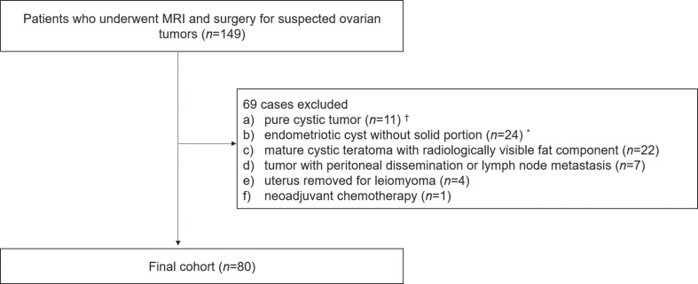
Methods: Data of 80 patients suspected of having adnexal tumors by ultrasonography between April 2008 and August 2018 were used for the study. All patients had undergone preoperative MRI and surgical resection at our institution. Four radiologists (two specialized in gynecological radiology and two non-specialized) were enrolled for blinded review of the MR images. A 3-point scale was used: 0 = benign, 1 = indeterminate, and 2 = malignant. Three imaging sets were reviewed: Set A, unenhanced MRI including DWI; Set B, Set A and delayed CE-T1WI; and Set C, Set A and DCE-MRI. Imaging criteria for benign and malignant tumors were given in earlier reports. The diagnostic performance of the three imaging sets of the four readers was calculated. Their areas under the curve (AUCs) were compared using the DeLong method.
Results: Accuracies of Set B were 81%-88%. Those of Set C were 81%-85%. The AUCs of Set B were 0.83 and 0.89. Those of Set C were 0.81-0.86. For two readers, Set A showed lower accuracy and AUC than Set B/Set C (less than 0.80), although those were equivalent in other readers. No significant difference in AUCs was found among the three sequence sets. Intrareader agreement was moderate to almost perfect in Sets A and B, and substantial to almost perfect in Set C.
Conclusion: DCE-MR showed no superiority for differentiating malignant adnexal tumors from benign tumors compared to delayed CE-T1WI with conventional MR and DWI.
期刊介绍:
Magnetic Resonance in Medical Sciences (MRMS or Magn
Reson Med Sci) is an international journal pursuing the
publication of original articles contributing to the progress
of magnetic resonance in the field of biomedical sciences
including technical developments and clinical applications.
MRMS is an official journal of the Japanese Society for
Magnetic Resonance in Medicine (JSMRM).


 分享
分享
 求助内容:
求助内容: 应助结果提醒方式:
 扫码关注我们
