Mala Naik, Morteza Esmaeili, Owen Thomas, Jonn T Geitung
{"title":"弥散张力成像是评估痴呆患者和行为问题并将其与其他痴呆患者区分开来的良好工具。","authors":"Mala Naik, Morteza Esmaeili, Owen Thomas, Jonn T Geitung","doi":"10.1177/20584601211066467","DOIUrl":null,"url":null,"abstract":"<p><strong>Background: </strong>Dementia is one of the leading public health concerns as the world's population ages. Although Alzheimer's disease (AD) is the most common dementia diagnosis among older patients, some patients have additional behavioral symptoms. It is therefore important to provide an exact diagnosis, both to provide the best possible treatment for patients and to facilitate better understanding.</p><p><strong>Purpose: </strong>To investigate whether magnetic resonance imaging (MRI) with fractional anisotropy (FA) can accurately find patients with behavioral symptoms within a group of AD patients.</p><p><strong>Material and methods: </strong>Forty-five patients from the geriatric outpatient clinic were recruited consecutively to form a group of patients with AD and behavioral symptoms (AD + BS) and a control group of 50 patients with established AD. All patients had a full assessment for dementia to establish the diagnosis according to ICD-10. MRI included 3D anatomical recordings for morphometric measurements, DTI for fiber tracking, and quantitative assessment of regional white matter integrity. The DTI analyses included computing of the diffusion tensor and its derived FA index.</p><p><strong>Results: </strong>We found a significant difference in FA values between the patient groups' frontal lobes. The FA was greater in the study group in both left (0.39 vs 0.09, <i>p</i> < 0.05) and right (0.40 vs 0.16, <i>p</i> < 0.05) frontal lobes.</p><p><strong>Conclusion: </strong>MRI with FA will find damage in frontal tracts and may be used as a diagnostic tool and be considered a robust tool for the recognizing different types of dementia in the future.</p>","PeriodicalId":72063,"journal":{"name":"Acta radiologica open","volume":"10 12","pages":"20584601211066467"},"PeriodicalIF":1.0000,"publicationDate":"2021-12-17","publicationTypes":"Journal Article","fieldsOfStudy":null,"isOpenAccess":false,"openAccessPdf":"https://www.ncbi.nlm.nih.gov/pmc/articles/PMC8689627/pdf/","citationCount":"0","resultStr":"{\"title\":\"Diffusion tension imaging is a good tool for assessing patients with dementia and behavioral problems and discriminating them from other dementia patients.\",\"authors\":\"Mala Naik, Morteza Esmaeili, Owen Thomas, Jonn T Geitung\",\"doi\":\"10.1177/20584601211066467\",\"DOIUrl\":null,\"url\":null,\"abstract\":\"<p><strong>Background: </strong>Dementia is one of the leading public health concerns as the world's population ages. Although Alzheimer's disease (AD) is the most common dementia diagnosis among older patients, some patients have additional behavioral symptoms. It is therefore important to provide an exact diagnosis, both to provide the best possible treatment for patients and to facilitate better understanding.</p><p><strong>Purpose: </strong>To investigate whether magnetic resonance imaging (MRI) with fractional anisotropy (FA) can accurately find patients with behavioral symptoms within a group of AD patients.</p><p><strong>Material and methods: </strong>Forty-five patients from the geriatric outpatient clinic were recruited consecutively to form a group of patients with AD and behavioral symptoms (AD + BS) and a control group of 50 patients with established AD. All patients had a full assessment for dementia to establish the diagnosis according to ICD-10. MRI included 3D anatomical recordings for morphometric measurements, DTI for fiber tracking, and quantitative assessment of regional white matter integrity. The DTI analyses included computing of the diffusion tensor and its derived FA index.</p><p><strong>Results: </strong>We found a significant difference in FA values between the patient groups' frontal lobes. The FA was greater in the study group in both left (0.39 vs 0.09, <i>p</i> < 0.05) and right (0.40 vs 0.16, <i>p</i> < 0.05) frontal lobes.</p><p><strong>Conclusion: </strong>MRI with FA will find damage in frontal tracts and may be used as a diagnostic tool and be considered a robust tool for the recognizing different types of dementia in the future.</p>\",\"PeriodicalId\":72063,\"journal\":{\"name\":\"Acta radiologica open\",\"volume\":\"10 12\",\"pages\":\"20584601211066467\"},\"PeriodicalIF\":1.0000,\"publicationDate\":\"2021-12-17\",\"publicationTypes\":\"Journal Article\",\"fieldsOfStudy\":null,\"isOpenAccess\":false,\"openAccessPdf\":\"https://www.ncbi.nlm.nih.gov/pmc/articles/PMC8689627/pdf/\",\"citationCount\":\"0\",\"resultStr\":null,\"platform\":\"Semanticscholar\",\"paperid\":null,\"PeriodicalName\":\"Acta radiologica open\",\"FirstCategoryId\":\"1085\",\"ListUrlMain\":\"https://doi.org/10.1177/20584601211066467\",\"RegionNum\":0,\"RegionCategory\":null,\"ArticlePicture\":[],\"TitleCN\":null,\"AbstractTextCN\":null,\"PMCID\":null,\"EPubDate\":\"2021/12/1 0:00:00\",\"PubModel\":\"eCollection\",\"JCR\":\"Q4\",\"JCRName\":\"RADIOLOGY, NUCLEAR MEDICINE & MEDICAL IMAGING\",\"Score\":null,\"Total\":0}","platform":"Semanticscholar","paperid":null,"PeriodicalName":"Acta radiologica open","FirstCategoryId":"1085","ListUrlMain":"https://doi.org/10.1177/20584601211066467","RegionNum":0,"RegionCategory":null,"ArticlePicture":[],"TitleCN":null,"AbstractTextCN":null,"PMCID":null,"EPubDate":"2021/12/1 0:00:00","PubModel":"eCollection","JCR":"Q4","JCRName":"RADIOLOGY, NUCLEAR MEDICINE & MEDICAL IMAGING","Score":null,"Total":0}
引用次数: 0
摘要
背景:随着世界人口老龄化,痴呆症是主要的公共卫生问题之一。虽然阿尔茨海默病(AD)是老年患者中最常见的痴呆症诊断,但一些患者有额外的行为症状。因此,提供准确的诊断非常重要,既可以为患者提供最好的治疗,也可以促进更好的理解。目的:探讨分数各向异性(FA)磁共振成像(MRI)能否准确发现AD患者组中存在行为症状的患者。材料与方法:从老年门诊连续招募45例AD伴行为症状患者(AD + BS)和50例已确诊AD患者作为对照组。所有患者都进行了全面的痴呆评估,以根据ICD-10确定诊断。MRI包括用于形态测量的3D解剖记录,用于纤维跟踪的DTI,以及区域白质完整性的定量评估。DTI分析包括计算扩散张量及其衍生的FA指数。结果:我们发现两组患者额叶FA值有显著差异。研究组左额叶FA (0.39 vs 0.09, p < 0.05)和右额叶FA (0.40 vs 0.16, p < 0.05)均高于对照组。结论:FA MRI可以发现额叶损伤,可作为一种诊断工具,并被认为是未来识别不同类型痴呆的有力工具。
Diffusion tension imaging is a good tool for assessing patients with dementia and behavioral problems and discriminating them from other dementia patients.
Background: Dementia is one of the leading public health concerns as the world's population ages. Although Alzheimer's disease (AD) is the most common dementia diagnosis among older patients, some patients have additional behavioral symptoms. It is therefore important to provide an exact diagnosis, both to provide the best possible treatment for patients and to facilitate better understanding.
Purpose: To investigate whether magnetic resonance imaging (MRI) with fractional anisotropy (FA) can accurately find patients with behavioral symptoms within a group of AD patients.
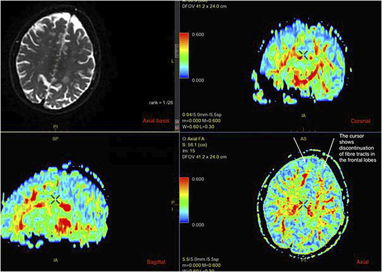
Material and methods: Forty-five patients from the geriatric outpatient clinic were recruited consecutively to form a group of patients with AD and behavioral symptoms (AD + BS) and a control group of 50 patients with established AD. All patients had a full assessment for dementia to establish the diagnosis according to ICD-10. MRI included 3D anatomical recordings for morphometric measurements, DTI for fiber tracking, and quantitative assessment of regional white matter integrity. The DTI analyses included computing of the diffusion tensor and its derived FA index.
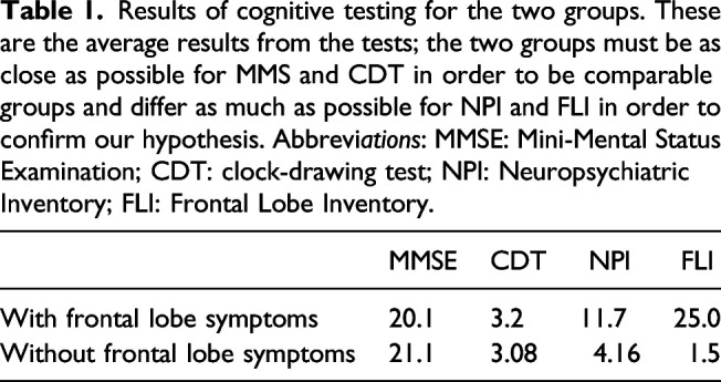
Results: We found a significant difference in FA values between the patient groups' frontal lobes. The FA was greater in the study group in both left (0.39 vs 0.09, p < 0.05) and right (0.40 vs 0.16, p < 0.05) frontal lobes.
Conclusion: MRI with FA will find damage in frontal tracts and may be used as a diagnostic tool and be considered a robust tool for the recognizing different types of dementia in the future.


 分享
分享

 求助内容:
求助内容: 应助结果提醒方式:
 扫码关注我们
