{"title":"腮腺有症状的插层导管病变:附免疫组织化学及基因分析1例。","authors":"Kimihide Kusafuka, Satoshi Baba, Yoshiharu Kitani, Kazuki Hirata, Akinori Murakami, Aya Muramatsu, Kazumori Arai, Makoto Suzuki","doi":"10.1007/s00795-022-00328-7","DOIUrl":null,"url":null,"abstract":"<p><p>Intercalated duct lesions (IDLs) are usually asymptomatic. We report a case of IDL, in which a palpable mass formed. The patient was a 45-year-old Japanese male, who noticed a mass in the left parotid region. The nodular lesion was well-circumscribed, but did not have a fibrous capsule or exhibit infiltrative growth. It contained a small cystic space and consisted of basaloid cells arranged in a cribriform pattern and inner ductal cells. It had some solid areas of nest-like proliferation displaying mild cellular atypia. Immunohistochemically, the luminal cells were positive for cytokeratin (CK)7 and epithelial membrane antigen, and the abluminal cells were positive for CK5/6, p63, and DOG1. S-100 protein-positive stromal cells were also seen. The lesion's cells were all positive for SOX10, and the nuclei of some basaloid cells were positive for β-catenin. The Ki-67 labeling index was 3.8%. The ductal cells contained diastase-digestion-resistant, Periodic acid Schiff-positive zymogen granules. Genetically, the lesion harbored a missense mutation in the CTNNB1 gene. We diagnosed the lesion as an IDL. As IDLs are usually small non-neoplastic lesions, symptomatic cases are rare. Based on its common immunohistochemical and genetic features, IDL may be a precursor of basal cell adenoma/adenocarcinoma, such as intercalated duct adenoma.</p>","PeriodicalId":18338,"journal":{"name":"Medical Molecular Morphology","volume":"55 4","pages":"329-336"},"PeriodicalIF":1.2000,"publicationDate":"2022-12-01","publicationTypes":"Journal Article","fieldsOfStudy":null,"isOpenAccess":false,"openAccessPdf":"","citationCount":"4","resultStr":"{\"title\":\"A symptomatic intercalated duct lesion of the parotid gland: a case report with immunohistochemical and genetic analyses.\",\"authors\":\"Kimihide Kusafuka, Satoshi Baba, Yoshiharu Kitani, Kazuki Hirata, Akinori Murakami, Aya Muramatsu, Kazumori Arai, Makoto Suzuki\",\"doi\":\"10.1007/s00795-022-00328-7\",\"DOIUrl\":null,\"url\":null,\"abstract\":\"<p><p>Intercalated duct lesions (IDLs) are usually asymptomatic. We report a case of IDL, in which a palpable mass formed. The patient was a 45-year-old Japanese male, who noticed a mass in the left parotid region. The nodular lesion was well-circumscribed, but did not have a fibrous capsule or exhibit infiltrative growth. It contained a small cystic space and consisted of basaloid cells arranged in a cribriform pattern and inner ductal cells. It had some solid areas of nest-like proliferation displaying mild cellular atypia. Immunohistochemically, the luminal cells were positive for cytokeratin (CK)7 and epithelial membrane antigen, and the abluminal cells were positive for CK5/6, p63, and DOG1. S-100 protein-positive stromal cells were also seen. The lesion's cells were all positive for SOX10, and the nuclei of some basaloid cells were positive for β-catenin. The Ki-67 labeling index was 3.8%. The ductal cells contained diastase-digestion-resistant, Periodic acid Schiff-positive zymogen granules. Genetically, the lesion harbored a missense mutation in the CTNNB1 gene. We diagnosed the lesion as an IDL. As IDLs are usually small non-neoplastic lesions, symptomatic cases are rare. Based on its common immunohistochemical and genetic features, IDL may be a precursor of basal cell adenoma/adenocarcinoma, such as intercalated duct adenoma.</p>\",\"PeriodicalId\":18338,\"journal\":{\"name\":\"Medical Molecular Morphology\",\"volume\":\"55 4\",\"pages\":\"329-336\"},\"PeriodicalIF\":1.2000,\"publicationDate\":\"2022-12-01\",\"publicationTypes\":\"Journal Article\",\"fieldsOfStudy\":null,\"isOpenAccess\":false,\"openAccessPdf\":\"\",\"citationCount\":\"4\",\"resultStr\":null,\"platform\":\"Semanticscholar\",\"paperid\":null,\"PeriodicalName\":\"Medical Molecular Morphology\",\"FirstCategoryId\":\"3\",\"ListUrlMain\":\"https://doi.org/10.1007/s00795-022-00328-7\",\"RegionNum\":4,\"RegionCategory\":\"医学\",\"ArticlePicture\":[],\"TitleCN\":null,\"AbstractTextCN\":null,\"PMCID\":null,\"EPubDate\":\"2022/7/5 0:00:00\",\"PubModel\":\"Epub\",\"JCR\":\"Q3\",\"JCRName\":\"PATHOLOGY\",\"Score\":null,\"Total\":0}","platform":"Semanticscholar","paperid":null,"PeriodicalName":"Medical Molecular Morphology","FirstCategoryId":"3","ListUrlMain":"https://doi.org/10.1007/s00795-022-00328-7","RegionNum":4,"RegionCategory":"医学","ArticlePicture":[],"TitleCN":null,"AbstractTextCN":null,"PMCID":null,"EPubDate":"2022/7/5 0:00:00","PubModel":"Epub","JCR":"Q3","JCRName":"PATHOLOGY","Score":null,"Total":0}
A symptomatic intercalated duct lesion of the parotid gland: a case report with immunohistochemical and genetic analyses.
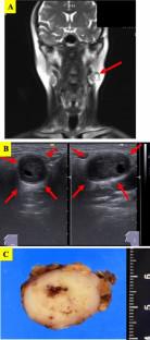
Intercalated duct lesions (IDLs) are usually asymptomatic. We report a case of IDL, in which a palpable mass formed. The patient was a 45-year-old Japanese male, who noticed a mass in the left parotid region. The nodular lesion was well-circumscribed, but did not have a fibrous capsule or exhibit infiltrative growth. It contained a small cystic space and consisted of basaloid cells arranged in a cribriform pattern and inner ductal cells. It had some solid areas of nest-like proliferation displaying mild cellular atypia. Immunohistochemically, the luminal cells were positive for cytokeratin (CK)7 and epithelial membrane antigen, and the abluminal cells were positive for CK5/6, p63, and DOG1. S-100 protein-positive stromal cells were also seen. The lesion's cells were all positive for SOX10, and the nuclei of some basaloid cells were positive for β-catenin. The Ki-67 labeling index was 3.8%. The ductal cells contained diastase-digestion-resistant, Periodic acid Schiff-positive zymogen granules. Genetically, the lesion harbored a missense mutation in the CTNNB1 gene. We diagnosed the lesion as an IDL. As IDLs are usually small non-neoplastic lesions, symptomatic cases are rare. Based on its common immunohistochemical and genetic features, IDL may be a precursor of basal cell adenoma/adenocarcinoma, such as intercalated duct adenoma.
期刊介绍:
Medical Molecular Morphology is an international forum for researchers in both basic and clinical medicine to present and discuss new research on the structural mechanisms and the processes of health and disease at the molecular level. The structures of molecules, organelles, cells, tissues, and organs determine their normal function. Disease is thus best understood in terms of structural changes in these different levels of biological organization, especially in molecules and molecular interactions as well as the cellular localization of chemical components. Medical Molecular Morphology welcomes articles on basic or clinical research in the fields of cell biology, molecular biology, and medical, veterinary, and dental sciences using techniques for structural research such as electron microscopy, confocal laser scanning microscopy, enzyme histochemistry, immunohistochemistry, radioautography, X-ray microanalysis, and in situ hybridization.
Manuscripts submitted for publication must contain a statement to the effect that all human studies have been reviewed by the appropriate ethics committee and have therefore been performed in accordance with the ethical standards laid down in an appropriate version of the 1964 Declaration of Helsinki. It should also be stated clearly in the text that all persons gave their informed consent prior to their inclusion in the study. Details that might disclose the identity of the subjects under study should be omitted.


 分享
分享
 求助内容:
求助内容: 应助结果提醒方式:
 扫码关注我们
