{"title":"在胰腺的自由呼吸动态对比增强MR成像中实现良好图像质量和进行药代动力学分析的最佳时间分辨率。","authors":"Kazuki Oyama, Fumihito Ichinohe, Akira Yamada, Yoshihiro Kitoh, Yasuo Adachi, Hayato Hayashihara, Marcel D Nickel, Katsuya Maruyama, Yasunari Fujinaga","doi":"10.2463/mrms.mp.2022-0035","DOIUrl":null,"url":null,"abstract":"<p><strong>Purpose: </strong>The optimal temporal resolution for free-breathing dynamic contrast-enhanced MRI (FBDCE-MRI) of the pancreas has not been determined. This study aimed to evaluate the appropriate temporal resolution to achieve good image quality and to perform pharmacokinetic analysis in FBDCE-MRI of the pancreas using golden-angle radial sparse parallel (GRASP).</p><p><strong>Methods: </strong>Sixteen participants (53 ± 15 years, eight females) undergoing FBDCE-MRI were included in this prospective study. Images were retrospectively reconstructed at four temporal resolutions (1.8, 3.0, 4.8, and 7.8s). Two radiologists (5 years of experience) evaluated the image quality of each reconstructed image by assessing the visualization of the celiac artery (CEA), the common hepatic artery, the splenic artery, each area of the pancreas, and artifacts using a 5-point scale. Using Tissue-4D, pharmacokinetic parameters were calculated for each area in the reconstructed images at each temporal resolution for 16 examinations, excluding two with errors in the pharmacokinetic modeling analysis. Friedman and Bonferroni tests were used for analysis. A P value < 0.05 was considered statistically significant.</p><p><strong>Results: </strong>During vascular assessment, only scores for the CEA at 7.8s were significantly lower than the other temporal resolutions. Scores of all pancreatic regions and artifacts were significantly lower at 1.8s than at 4.8s and 7.8s. In the pharmacokinetic analysis, all volume transfer coefficients (K<sub>trans</sub>), rate constants (K<sub>ep</sub>), and the initial area under the concentration curve (iAUC) in the pancreatic head and tail were significantly lower at 4.8s and 7.8s than at 1.8s. iAUC in the pancreatic body and extracellular extravascular volume fraction (V<sub>e</sub>) in the pancreatic head were significantly lower at 7.8s than at 1.8s.</p><p><strong>Conclusion: </strong>A temporal resolution of 3.0s is appropriate to achieve image quality and perform pharmacokinetic analysis in FBDCE-MRI of the pancreas using GRASP.</p>","PeriodicalId":2,"journal":{"name":"ACS Applied Bio Materials","volume":" ","pages":"477-485"},"PeriodicalIF":4.7000,"publicationDate":"2023-10-01","publicationTypes":"Journal Article","fieldsOfStudy":null,"isOpenAccess":false,"openAccessPdf":"https://ftp.ncbi.nlm.nih.gov/pub/pmc/oa_pdf/a1/9f/mrms-22-477.PMC10552666.pdf","citationCount":"1","resultStr":"{\"title\":\"Optimal Temporal Resolution to Achieve Good Image Quality and Perform Pharmacokinetic Analysis in Free-breathing Dynamic Contrast-enhanced MR Imaging of the Pancreas.\",\"authors\":\"Kazuki Oyama, Fumihito Ichinohe, Akira Yamada, Yoshihiro Kitoh, Yasuo Adachi, Hayato Hayashihara, Marcel D Nickel, Katsuya Maruyama, Yasunari Fujinaga\",\"doi\":\"10.2463/mrms.mp.2022-0035\",\"DOIUrl\":null,\"url\":null,\"abstract\":\"<p><strong>Purpose: </strong>The optimal temporal resolution for free-breathing dynamic contrast-enhanced MRI (FBDCE-MRI) of the pancreas has not been determined. This study aimed to evaluate the appropriate temporal resolution to achieve good image quality and to perform pharmacokinetic analysis in FBDCE-MRI of the pancreas using golden-angle radial sparse parallel (GRASP).</p><p><strong>Methods: </strong>Sixteen participants (53 ± 15 years, eight females) undergoing FBDCE-MRI were included in this prospective study. Images were retrospectively reconstructed at four temporal resolutions (1.8, 3.0, 4.8, and 7.8s). Two radiologists (5 years of experience) evaluated the image quality of each reconstructed image by assessing the visualization of the celiac artery (CEA), the common hepatic artery, the splenic artery, each area of the pancreas, and artifacts using a 5-point scale. Using Tissue-4D, pharmacokinetic parameters were calculated for each area in the reconstructed images at each temporal resolution for 16 examinations, excluding two with errors in the pharmacokinetic modeling analysis. Friedman and Bonferroni tests were used for analysis. A P value < 0.05 was considered statistically significant.</p><p><strong>Results: </strong>During vascular assessment, only scores for the CEA at 7.8s were significantly lower than the other temporal resolutions. Scores of all pancreatic regions and artifacts were significantly lower at 1.8s than at 4.8s and 7.8s. In the pharmacokinetic analysis, all volume transfer coefficients (K<sub>trans</sub>), rate constants (K<sub>ep</sub>), and the initial area under the concentration curve (iAUC) in the pancreatic head and tail were significantly lower at 4.8s and 7.8s than at 1.8s. iAUC in the pancreatic body and extracellular extravascular volume fraction (V<sub>e</sub>) in the pancreatic head were significantly lower at 7.8s than at 1.8s.</p><p><strong>Conclusion: </strong>A temporal resolution of 3.0s is appropriate to achieve image quality and perform pharmacokinetic analysis in FBDCE-MRI of the pancreas using GRASP.</p>\",\"PeriodicalId\":2,\"journal\":{\"name\":\"ACS Applied Bio Materials\",\"volume\":\" \",\"pages\":\"477-485\"},\"PeriodicalIF\":4.7000,\"publicationDate\":\"2023-10-01\",\"publicationTypes\":\"Journal Article\",\"fieldsOfStudy\":null,\"isOpenAccess\":false,\"openAccessPdf\":\"https://ftp.ncbi.nlm.nih.gov/pub/pmc/oa_pdf/a1/9f/mrms-22-477.PMC10552666.pdf\",\"citationCount\":\"1\",\"resultStr\":null,\"platform\":\"Semanticscholar\",\"paperid\":null,\"PeriodicalName\":\"ACS Applied Bio Materials\",\"FirstCategoryId\":\"3\",\"ListUrlMain\":\"https://doi.org/10.2463/mrms.mp.2022-0035\",\"RegionNum\":0,\"RegionCategory\":null,\"ArticlePicture\":[],\"TitleCN\":null,\"AbstractTextCN\":null,\"PMCID\":null,\"EPubDate\":\"2022/8/23 0:00:00\",\"PubModel\":\"Epub\",\"JCR\":\"Q2\",\"JCRName\":\"MATERIALS SCIENCE, BIOMATERIALS\",\"Score\":null,\"Total\":0}","platform":"Semanticscholar","paperid":null,"PeriodicalName":"ACS Applied Bio Materials","FirstCategoryId":"3","ListUrlMain":"https://doi.org/10.2463/mrms.mp.2022-0035","RegionNum":0,"RegionCategory":null,"ArticlePicture":[],"TitleCN":null,"AbstractTextCN":null,"PMCID":null,"EPubDate":"2022/8/23 0:00:00","PubModel":"Epub","JCR":"Q2","JCRName":"MATERIALS SCIENCE, BIOMATERIALS","Score":null,"Total":0}
Optimal Temporal Resolution to Achieve Good Image Quality and Perform Pharmacokinetic Analysis in Free-breathing Dynamic Contrast-enhanced MR Imaging of the Pancreas.
Purpose: The optimal temporal resolution for free-breathing dynamic contrast-enhanced MRI (FBDCE-MRI) of the pancreas has not been determined. This study aimed to evaluate the appropriate temporal resolution to achieve good image quality and to perform pharmacokinetic analysis in FBDCE-MRI of the pancreas using golden-angle radial sparse parallel (GRASP).
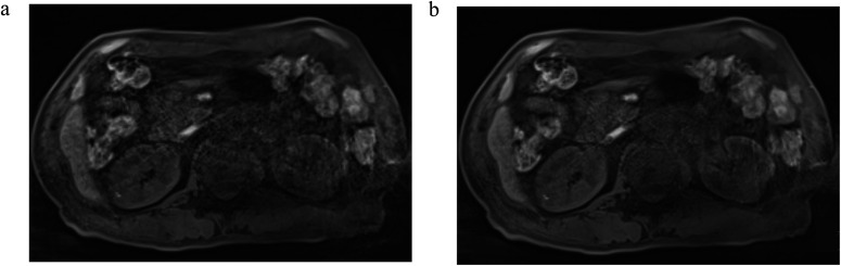
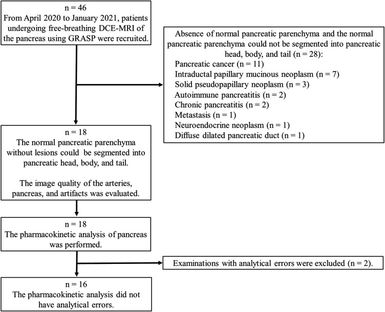
Methods: Sixteen participants (53 ± 15 years, eight females) undergoing FBDCE-MRI were included in this prospective study. Images were retrospectively reconstructed at four temporal resolutions (1.8, 3.0, 4.8, and 7.8s). Two radiologists (5 years of experience) evaluated the image quality of each reconstructed image by assessing the visualization of the celiac artery (CEA), the common hepatic artery, the splenic artery, each area of the pancreas, and artifacts using a 5-point scale. Using Tissue-4D, pharmacokinetic parameters were calculated for each area in the reconstructed images at each temporal resolution for 16 examinations, excluding two with errors in the pharmacokinetic modeling analysis. Friedman and Bonferroni tests were used for analysis. A P value < 0.05 was considered statistically significant.
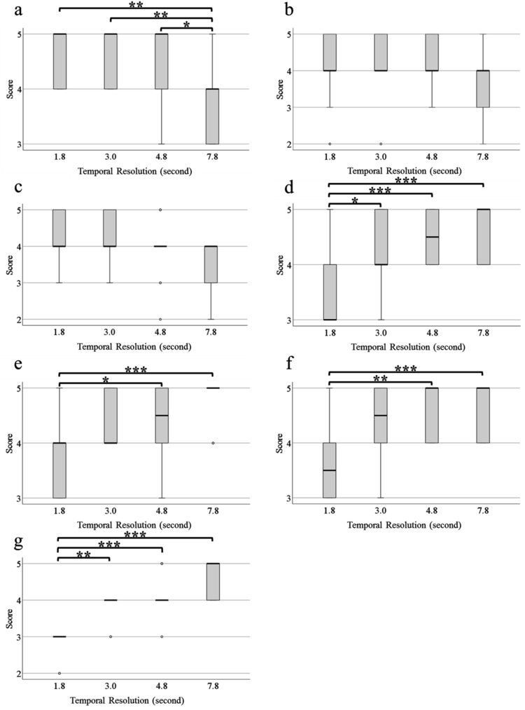
Results: During vascular assessment, only scores for the CEA at 7.8s were significantly lower than the other temporal resolutions. Scores of all pancreatic regions and artifacts were significantly lower at 1.8s than at 4.8s and 7.8s. In the pharmacokinetic analysis, all volume transfer coefficients (Ktrans), rate constants (Kep), and the initial area under the concentration curve (iAUC) in the pancreatic head and tail were significantly lower at 4.8s and 7.8s than at 1.8s. iAUC in the pancreatic body and extracellular extravascular volume fraction (Ve) in the pancreatic head were significantly lower at 7.8s than at 1.8s.
Conclusion: A temporal resolution of 3.0s is appropriate to achieve image quality and perform pharmacokinetic analysis in FBDCE-MRI of the pancreas using GRASP.
期刊介绍:
ACS Applied Bio Materials is an interdisciplinary journal publishing original research covering all aspects of biomaterials and biointerfaces including and beyond the traditional biosensing, biomedical and therapeutic applications.
The journal is devoted to reports of new and original experimental and theoretical research of an applied nature that integrates knowledge in the areas of materials, engineering, physics, bioscience, and chemistry into important bio applications. The journal is specifically interested in work that addresses the relationship between structure and function and assesses the stability and degradation of materials under relevant environmental and biological conditions.


 分享
分享
 求助内容:
求助内容: 应助结果提醒方式:
 扫码关注我们
