Blake H Fortes, Aaron M Fairbanks, Aravindh A Nirmalan, David O Hodge, Kevin Ferenchak, Andrew J Barkmeier
{"title":"渗出性年龄相关性黄斑变性中基于光学相干断层扫描的黄斑液的日变化。","authors":"Blake H Fortes, Aaron M Fairbanks, Aravindh A Nirmalan, David O Hodge, Kevin Ferenchak, Andrew J Barkmeier","doi":"10.1186/s40942-023-00495-4","DOIUrl":null,"url":null,"abstract":"<p><strong>Background: </strong>Significant diurnal fluctuation of optical coherence tomography (OCT)-based macular fluid occurs in patients with several macular conditions including diabetic macular edema (DME) and cystoid macular edema due to retinal venous occlusion (RVO). OCT imaging and analysis of macular fluid status plays a central role in clinical management of exudative age-related macular degeneration (eAMD), however diurnal variation of eAMD OCT findings has not yet been formally studied. Herein, we investigate whether clinically meaningful fluctuation of OCT-based macular fluid occurs in patients with eAMD.</p><p><strong>Methods: </strong>Prospective observational study. Patients with eAMD and intra- and/or sub-retinal fluid on early AM OCT were enrolled to receive two consecutive OCT scans at least four hours later. Retinal layers were manually segmented on all OCT rasters and AM-to-PM and PM-to-PM image pairs were analyzed for total retinal and neurosensory retinal volume changes within the central 1 and 3 mm ETDRS subfields. Finally, two masked retinal specialists analyzed all OCT image pairs for qualitative differences that may impact clinical management.</p><p><strong>Results: </strong>21 patients with eAMD and fluid on OCT were recruited between January 2020 and November 2021. There was no mean difference between AM and PM central 3 mm total retinal volume (p = 0.56), central 3 mm neurosensory retinal volume (p = 0.25), central 1 mm total retinal mean thickness (p = 0.96), or central 1 mm neurosensory retinal mean thickness (p = 0.63), nor were any differences identified in PM-to-PM control comparisons. Qualitative analysis by two masked experts identified no clinically significant differences between any AM-to-PM OCT image pairs.</p><p><strong>Conclusions: </strong>No significant diurnal variation in OCT-based macular fluid or thickness was identified in patients with eAMD, either quantitatively or qualitatively.</p>","PeriodicalId":14289,"journal":{"name":"International Journal of Retina and Vitreous","volume":null,"pages":null},"PeriodicalIF":1.9000,"publicationDate":"2023-09-25","publicationTypes":"Journal Article","fieldsOfStudy":null,"isOpenAccess":false,"openAccessPdf":"https://www.ncbi.nlm.nih.gov/pmc/articles/PMC10518912/pdf/","citationCount":"0","resultStr":"{\"title\":\"Diurnal variation of optical coherence tomography-based macular fluid in exudative age-related macular degeneration.\",\"authors\":\"Blake H Fortes, Aaron M Fairbanks, Aravindh A Nirmalan, David O Hodge, Kevin Ferenchak, Andrew J Barkmeier\",\"doi\":\"10.1186/s40942-023-00495-4\",\"DOIUrl\":null,\"url\":null,\"abstract\":\"<p><strong>Background: </strong>Significant diurnal fluctuation of optical coherence tomography (OCT)-based macular fluid occurs in patients with several macular conditions including diabetic macular edema (DME) and cystoid macular edema due to retinal venous occlusion (RVO). OCT imaging and analysis of macular fluid status plays a central role in clinical management of exudative age-related macular degeneration (eAMD), however diurnal variation of eAMD OCT findings has not yet been formally studied. Herein, we investigate whether clinically meaningful fluctuation of OCT-based macular fluid occurs in patients with eAMD.</p><p><strong>Methods: </strong>Prospective observational study. Patients with eAMD and intra- and/or sub-retinal fluid on early AM OCT were enrolled to receive two consecutive OCT scans at least four hours later. Retinal layers were manually segmented on all OCT rasters and AM-to-PM and PM-to-PM image pairs were analyzed for total retinal and neurosensory retinal volume changes within the central 1 and 3 mm ETDRS subfields. Finally, two masked retinal specialists analyzed all OCT image pairs for qualitative differences that may impact clinical management.</p><p><strong>Results: </strong>21 patients with eAMD and fluid on OCT were recruited between January 2020 and November 2021. There was no mean difference between AM and PM central 3 mm total retinal volume (p = 0.56), central 3 mm neurosensory retinal volume (p = 0.25), central 1 mm total retinal mean thickness (p = 0.96), or central 1 mm neurosensory retinal mean thickness (p = 0.63), nor were any differences identified in PM-to-PM control comparisons. Qualitative analysis by two masked experts identified no clinically significant differences between any AM-to-PM OCT image pairs.</p><p><strong>Conclusions: </strong>No significant diurnal variation in OCT-based macular fluid or thickness was identified in patients with eAMD, either quantitatively or qualitatively.</p>\",\"PeriodicalId\":14289,\"journal\":{\"name\":\"International Journal of Retina and Vitreous\",\"volume\":null,\"pages\":null},\"PeriodicalIF\":1.9000,\"publicationDate\":\"2023-09-25\",\"publicationTypes\":\"Journal Article\",\"fieldsOfStudy\":null,\"isOpenAccess\":false,\"openAccessPdf\":\"https://www.ncbi.nlm.nih.gov/pmc/articles/PMC10518912/pdf/\",\"citationCount\":\"0\",\"resultStr\":null,\"platform\":\"Semanticscholar\",\"paperid\":null,\"PeriodicalName\":\"International Journal of Retina and Vitreous\",\"FirstCategoryId\":\"1085\",\"ListUrlMain\":\"https://doi.org/10.1186/s40942-023-00495-4\",\"RegionNum\":0,\"RegionCategory\":null,\"ArticlePicture\":[],\"TitleCN\":null,\"AbstractTextCN\":null,\"PMCID\":null,\"EPubDate\":\"\",\"PubModel\":\"\",\"JCR\":\"Q2\",\"JCRName\":\"OPHTHALMOLOGY\",\"Score\":null,\"Total\":0}","platform":"Semanticscholar","paperid":null,"PeriodicalName":"International Journal of Retina and Vitreous","FirstCategoryId":"1085","ListUrlMain":"https://doi.org/10.1186/s40942-023-00495-4","RegionNum":0,"RegionCategory":null,"ArticlePicture":[],"TitleCN":null,"AbstractTextCN":null,"PMCID":null,"EPubDate":"","PubModel":"","JCR":"Q2","JCRName":"OPHTHALMOLOGY","Score":null,"Total":0}
Diurnal variation of optical coherence tomography-based macular fluid in exudative age-related macular degeneration.
Background: Significant diurnal fluctuation of optical coherence tomography (OCT)-based macular fluid occurs in patients with several macular conditions including diabetic macular edema (DME) and cystoid macular edema due to retinal venous occlusion (RVO). OCT imaging and analysis of macular fluid status plays a central role in clinical management of exudative age-related macular degeneration (eAMD), however diurnal variation of eAMD OCT findings has not yet been formally studied. Herein, we investigate whether clinically meaningful fluctuation of OCT-based macular fluid occurs in patients with eAMD.
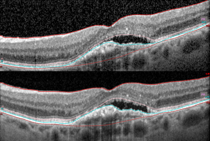
Methods: Prospective observational study. Patients with eAMD and intra- and/or sub-retinal fluid on early AM OCT were enrolled to receive two consecutive OCT scans at least four hours later. Retinal layers were manually segmented on all OCT rasters and AM-to-PM and PM-to-PM image pairs were analyzed for total retinal and neurosensory retinal volume changes within the central 1 and 3 mm ETDRS subfields. Finally, two masked retinal specialists analyzed all OCT image pairs for qualitative differences that may impact clinical management.
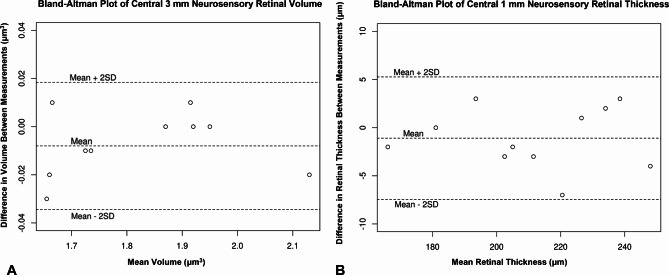
Results: 21 patients with eAMD and fluid on OCT were recruited between January 2020 and November 2021. There was no mean difference between AM and PM central 3 mm total retinal volume (p = 0.56), central 3 mm neurosensory retinal volume (p = 0.25), central 1 mm total retinal mean thickness (p = 0.96), or central 1 mm neurosensory retinal mean thickness (p = 0.63), nor were any differences identified in PM-to-PM control comparisons. Qualitative analysis by two masked experts identified no clinically significant differences between any AM-to-PM OCT image pairs.
Conclusions: No significant diurnal variation in OCT-based macular fluid or thickness was identified in patients with eAMD, either quantitatively or qualitatively.
期刊介绍:
International Journal of Retina and Vitreous focuses on the ophthalmic subspecialty of vitreoretinal disorders. The journal presents original articles on new approaches to diagnosis, outcomes of clinical trials, innovations in pharmacological therapy and surgical techniques, as well as basic science advances that impact clinical practice. Topical areas include, but are not limited to: -Imaging of the retina, choroid and vitreous -Innovations in optical coherence tomography (OCT) -Small-gauge vitrectomy, retinal detachment, chromovitrectomy -Electroretinography (ERG), microperimetry, other functional tests -Intraocular tumors -Retinal pharmacotherapy & drug delivery -Diabetic retinopathy & other vascular diseases -Age-related macular degeneration (AMD) & other macular entities


 分享
分享
 求助内容:
求助内容: 应助结果提醒方式:
 扫码关注我们
