Gabriela Dziurman, Agnieszka Drzał, Aleksandra Anna Murzyn, Maciej Mikolaj Kmiec, Martyna Elas, Martyna Krzykawska-Serda
{"title":"在吉西他滨治疗的小鼠胰腺肿瘤中使用Oxychip的脉冲和CW EPR血氧测定。","authors":"Gabriela Dziurman, Agnieszka Drzał, Aleksandra Anna Murzyn, Maciej Mikolaj Kmiec, Martyna Elas, Martyna Krzykawska-Serda","doi":"10.1007/s11307-023-01859-w","DOIUrl":null,"url":null,"abstract":"<p><strong>Purpose: </strong>The goal of this work was to compare pO<sub>2</sub> measured using both continuous wave (CW) and pulse electron paramagnetic resonance (EPR) spectroscopy. The Oxychip particle spin probe enabled longitudinal monitoring of pO<sub>2</sub> in murine pancreatic tumor treated with gemcitabine during the course of therapy.</p><p><strong>Procedures: </strong>Pancreatic PanO2 tumors were growing in the syngeneic mice, in the leg. Five doses of saline in control animals or gemcitabine were administered every 3 days, and pO<sub>2</sub> was measured after each dose at several time points. Oxygen partial pressure was determined from the linewidth of the CW EPR signal (Bruker E540L) or from the T<sub>2</sub> measured using the electron spin echo sequence (Jiva-25™).</p><p><strong>Results: </strong>The oxygen sensitivity was determined from a calibration curve as 6.1 mG/mm Hg in CW EPR and 68.5 ms<sup>-1</sup>/mm Hg in pulse EPR. A slight increase in pO<sub>2</sub> of up to 20 mm Hg was observed after the third dose of gemcitabine compared to the control. The maximum delta pO<sub>2</sub> during the therapy correlated with better survival.</p><p><strong>Conclusions: </strong>Both techniques offer fast and reliable oximetry in vivo, allowing to follow the effects of pharmaceutic intervention.</p>","PeriodicalId":18760,"journal":{"name":"Molecular Imaging and Biology","volume":" ","pages":"473-483"},"PeriodicalIF":2.5000,"publicationDate":"2024-06-01","publicationTypes":"Journal Article","fieldsOfStudy":null,"isOpenAccess":false,"openAccessPdf":"https://www.ncbi.nlm.nih.gov/pmc/articles/PMC11211198/pdf/","citationCount":"0","resultStr":"{\"title\":\"Pulse and CW EPR Oximetry Using Oxychip in Gemcitabine-Treated Murine Pancreatic Tumors.\",\"authors\":\"Gabriela Dziurman, Agnieszka Drzał, Aleksandra Anna Murzyn, Maciej Mikolaj Kmiec, Martyna Elas, Martyna Krzykawska-Serda\",\"doi\":\"10.1007/s11307-023-01859-w\",\"DOIUrl\":null,\"url\":null,\"abstract\":\"<p><strong>Purpose: </strong>The goal of this work was to compare pO<sub>2</sub> measured using both continuous wave (CW) and pulse electron paramagnetic resonance (EPR) spectroscopy. The Oxychip particle spin probe enabled longitudinal monitoring of pO<sub>2</sub> in murine pancreatic tumor treated with gemcitabine during the course of therapy.</p><p><strong>Procedures: </strong>Pancreatic PanO2 tumors were growing in the syngeneic mice, in the leg. Five doses of saline in control animals or gemcitabine were administered every 3 days, and pO<sub>2</sub> was measured after each dose at several time points. Oxygen partial pressure was determined from the linewidth of the CW EPR signal (Bruker E540L) or from the T<sub>2</sub> measured using the electron spin echo sequence (Jiva-25™).</p><p><strong>Results: </strong>The oxygen sensitivity was determined from a calibration curve as 6.1 mG/mm Hg in CW EPR and 68.5 ms<sup>-1</sup>/mm Hg in pulse EPR. A slight increase in pO<sub>2</sub> of up to 20 mm Hg was observed after the third dose of gemcitabine compared to the control. The maximum delta pO<sub>2</sub> during the therapy correlated with better survival.</p><p><strong>Conclusions: </strong>Both techniques offer fast and reliable oximetry in vivo, allowing to follow the effects of pharmaceutic intervention.</p>\",\"PeriodicalId\":18760,\"journal\":{\"name\":\"Molecular Imaging and Biology\",\"volume\":\" \",\"pages\":\"473-483\"},\"PeriodicalIF\":2.5000,\"publicationDate\":\"2024-06-01\",\"publicationTypes\":\"Journal Article\",\"fieldsOfStudy\":null,\"isOpenAccess\":false,\"openAccessPdf\":\"https://www.ncbi.nlm.nih.gov/pmc/articles/PMC11211198/pdf/\",\"citationCount\":\"0\",\"resultStr\":null,\"platform\":\"Semanticscholar\",\"paperid\":null,\"PeriodicalName\":\"Molecular Imaging and Biology\",\"FirstCategoryId\":\"3\",\"ListUrlMain\":\"https://doi.org/10.1007/s11307-023-01859-w\",\"RegionNum\":4,\"RegionCategory\":\"医学\",\"ArticlePicture\":[],\"TitleCN\":null,\"AbstractTextCN\":null,\"PMCID\":null,\"EPubDate\":\"2023/10/2 0:00:00\",\"PubModel\":\"Epub\",\"JCR\":\"Q2\",\"JCRName\":\"RADIOLOGY, NUCLEAR MEDICINE & MEDICAL IMAGING\",\"Score\":null,\"Total\":0}","platform":"Semanticscholar","paperid":null,"PeriodicalName":"Molecular Imaging and Biology","FirstCategoryId":"3","ListUrlMain":"https://doi.org/10.1007/s11307-023-01859-w","RegionNum":4,"RegionCategory":"医学","ArticlePicture":[],"TitleCN":null,"AbstractTextCN":null,"PMCID":null,"EPubDate":"2023/10/2 0:00:00","PubModel":"Epub","JCR":"Q2","JCRName":"RADIOLOGY, NUCLEAR MEDICINE & MEDICAL IMAGING","Score":null,"Total":0}
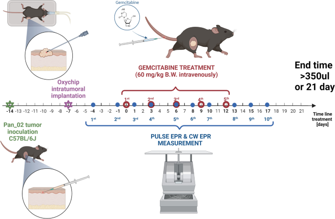
Pulse and CW EPR Oximetry Using Oxychip in Gemcitabine-Treated Murine Pancreatic Tumors.
Purpose: The goal of this work was to compare pO2 measured using both continuous wave (CW) and pulse electron paramagnetic resonance (EPR) spectroscopy. The Oxychip particle spin probe enabled longitudinal monitoring of pO2 in murine pancreatic tumor treated with gemcitabine during the course of therapy.
Procedures: Pancreatic PanO2 tumors were growing in the syngeneic mice, in the leg. Five doses of saline in control animals or gemcitabine were administered every 3 days, and pO2 was measured after each dose at several time points. Oxygen partial pressure was determined from the linewidth of the CW EPR signal (Bruker E540L) or from the T2 measured using the electron spin echo sequence (Jiva-25™).
Results: The oxygen sensitivity was determined from a calibration curve as 6.1 mG/mm Hg in CW EPR and 68.5 ms-1/mm Hg in pulse EPR. A slight increase in pO2 of up to 20 mm Hg was observed after the third dose of gemcitabine compared to the control. The maximum delta pO2 during the therapy correlated with better survival.
Conclusions: Both techniques offer fast and reliable oximetry in vivo, allowing to follow the effects of pharmaceutic intervention.
期刊介绍:
Molecular Imaging and Biology (MIB) invites original contributions (research articles, review articles, commentaries, etc.) on the utilization of molecular imaging (i.e., nuclear imaging, optical imaging, autoradiography and pathology, MRI, MPI, ultrasound imaging, radiomics/genomics etc.) to investigate questions related to biology and health. The objective of MIB is to provide a forum to the discovery of molecular mechanisms of disease through the use of imaging techniques. We aim to investigate the biological nature of disease in patients and establish new molecular imaging diagnostic and therapy procedures.
Some areas that are covered are:
Preclinical and clinical imaging of macromolecular targets (e.g., genes, receptors, enzymes) involved in significant biological processes.
The design, characterization, and study of new molecular imaging probes and contrast agents for the functional interrogation of macromolecular targets.
Development and evaluation of imaging systems including instrumentation, image reconstruction algorithms, image analysis, and display.
Development of molecular assay approaches leading to quantification of the biological information obtained in molecular imaging.
Study of in vivo animal models of disease for the development of new molecular diagnostics and therapeutics.
Extension of in vitro and in vivo discoveries using disease models, into well designed clinical research investigations.
Clinical molecular imaging involving clinical investigations, clinical trials and medical management or cost-effectiveness studies.


 分享
分享

 求助内容:
求助内容: 应助结果提醒方式:
 扫码关注我们
