George E Finney, Kerrie E Hargrave, Marieke Pingen, Thomas Purnell, David Todd, Freya MacDonald, Julie C Worrell, Megan K L MacLeod
{"title":"甲型流感病毒感染后先天性和适应性淋巴细胞产生IFNγ的三相性。","authors":"George E Finney, Kerrie E Hargrave, Marieke Pingen, Thomas Purnell, David Todd, Freya MacDonald, Julie C Worrell, Megan K L MacLeod","doi":"10.1093/discim/kyad014","DOIUrl":null,"url":null,"abstract":"<p><p>Interferon gamma (IFN<i>γ</i>) is a potent antiviral cytokine that can be produced by many innate and adaptive immune cells during infection. Currently, our understanding of which cells produce IFN<i>γ</i> and where they are located at different stages of an infection is limited. We have used reporter mice to investigate <i>in vivo</i> expression of <i>Ifn</i><i>γ</i> mRNA in the lung and secondary lymphoid organs during and following influenza A virus (IAV) infection. We observed a triphasic production of <i>Ifn</i><i>γ</i> expression. Unconventional T cells and innate lymphoid cells, particularly NK cells, were the dominant producers of early <i>Ifn</i><i>γ</i>, while CD4 and CD8 T cells were the main producers by day 10 post-infection. Following viral clearance, some memory CD4 and CD8 T cells continued to express <i>Ifn</i><i>γ</i> in the lungs and draining lymph node. Interestingly, <i>Ifn</i><i>γ</i> production by lymph node natural killer (NK), NKT, and innate lymphoid type 1 cells also continued to be above naïve levels, suggesting memory-like phenotypes for these cells. Analysis of the localization of <i>Ifn</i><i>γ</i>+ memory CD4 and CD8 T cells demonstrated that cytokine+ T cells were located near airways and in the lung parenchyma. Following a second IAV challenge, lung IAV-specific CD8 T cells rapidly increased their expression of <i>Ifn</i><i>γ</i> while CD4 T cells in the draining lymph node increased their <i>Ifn</i><i>γ</i> response. Together, these data suggest that <i>Ifn</i><i>γ</i> production fluctuates based on cellular source and location, both of which could impact subsequent immune responses.</p>","PeriodicalId":72830,"journal":{"name":"Discovery immunology","volume":"2 1","pages":"kyad014"},"PeriodicalIF":0.0000,"publicationDate":"2023-08-19","publicationTypes":"Journal Article","fieldsOfStudy":null,"isOpenAccess":false,"openAccessPdf":"https://www.ncbi.nlm.nih.gov/pmc/articles/PMC10568397/pdf/","citationCount":"0","resultStr":"{\"title\":\"Triphasic production of IFN<i>γ</i> by innate and adaptive lymphocytes following influenza A virus infection.\",\"authors\":\"George E Finney, Kerrie E Hargrave, Marieke Pingen, Thomas Purnell, David Todd, Freya MacDonald, Julie C Worrell, Megan K L MacLeod\",\"doi\":\"10.1093/discim/kyad014\",\"DOIUrl\":null,\"url\":null,\"abstract\":\"<p><p>Interferon gamma (IFN<i>γ</i>) is a potent antiviral cytokine that can be produced by many innate and adaptive immune cells during infection. Currently, our understanding of which cells produce IFN<i>γ</i> and where they are located at different stages of an infection is limited. We have used reporter mice to investigate <i>in vivo</i> expression of <i>Ifn</i><i>γ</i> mRNA in the lung and secondary lymphoid organs during and following influenza A virus (IAV) infection. We observed a triphasic production of <i>Ifn</i><i>γ</i> expression. Unconventional T cells and innate lymphoid cells, particularly NK cells, were the dominant producers of early <i>Ifn</i><i>γ</i>, while CD4 and CD8 T cells were the main producers by day 10 post-infection. Following viral clearance, some memory CD4 and CD8 T cells continued to express <i>Ifn</i><i>γ</i> in the lungs and draining lymph node. Interestingly, <i>Ifn</i><i>γ</i> production by lymph node natural killer (NK), NKT, and innate lymphoid type 1 cells also continued to be above naïve levels, suggesting memory-like phenotypes for these cells. Analysis of the localization of <i>Ifn</i><i>γ</i>+ memory CD4 and CD8 T cells demonstrated that cytokine+ T cells were located near airways and in the lung parenchyma. Following a second IAV challenge, lung IAV-specific CD8 T cells rapidly increased their expression of <i>Ifn</i><i>γ</i> while CD4 T cells in the draining lymph node increased their <i>Ifn</i><i>γ</i> response. Together, these data suggest that <i>Ifn</i><i>γ</i> production fluctuates based on cellular source and location, both of which could impact subsequent immune responses.</p>\",\"PeriodicalId\":72830,\"journal\":{\"name\":\"Discovery immunology\",\"volume\":\"2 1\",\"pages\":\"kyad014\"},\"PeriodicalIF\":0.0000,\"publicationDate\":\"2023-08-19\",\"publicationTypes\":\"Journal Article\",\"fieldsOfStudy\":null,\"isOpenAccess\":false,\"openAccessPdf\":\"https://www.ncbi.nlm.nih.gov/pmc/articles/PMC10568397/pdf/\",\"citationCount\":\"0\",\"resultStr\":null,\"platform\":\"Semanticscholar\",\"paperid\":null,\"PeriodicalName\":\"Discovery immunology\",\"FirstCategoryId\":\"1085\",\"ListUrlMain\":\"https://doi.org/10.1093/discim/kyad014\",\"RegionNum\":0,\"RegionCategory\":null,\"ArticlePicture\":[],\"TitleCN\":null,\"AbstractTextCN\":null,\"PMCID\":null,\"EPubDate\":\"2023/1/1 0:00:00\",\"PubModel\":\"eCollection\",\"JCR\":\"\",\"JCRName\":\"\",\"Score\":null,\"Total\":0}","platform":"Semanticscholar","paperid":null,"PeriodicalName":"Discovery immunology","FirstCategoryId":"1085","ListUrlMain":"https://doi.org/10.1093/discim/kyad014","RegionNum":0,"RegionCategory":null,"ArticlePicture":[],"TitleCN":null,"AbstractTextCN":null,"PMCID":null,"EPubDate":"2023/1/1 0:00:00","PubModel":"eCollection","JCR":"","JCRName":"","Score":null,"Total":0}
Triphasic production of IFNγ by innate and adaptive lymphocytes following influenza A virus infection.
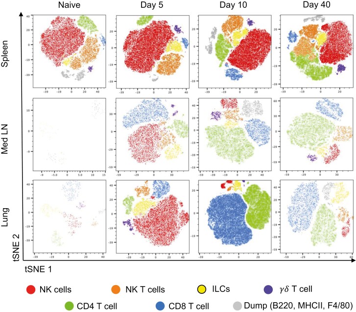
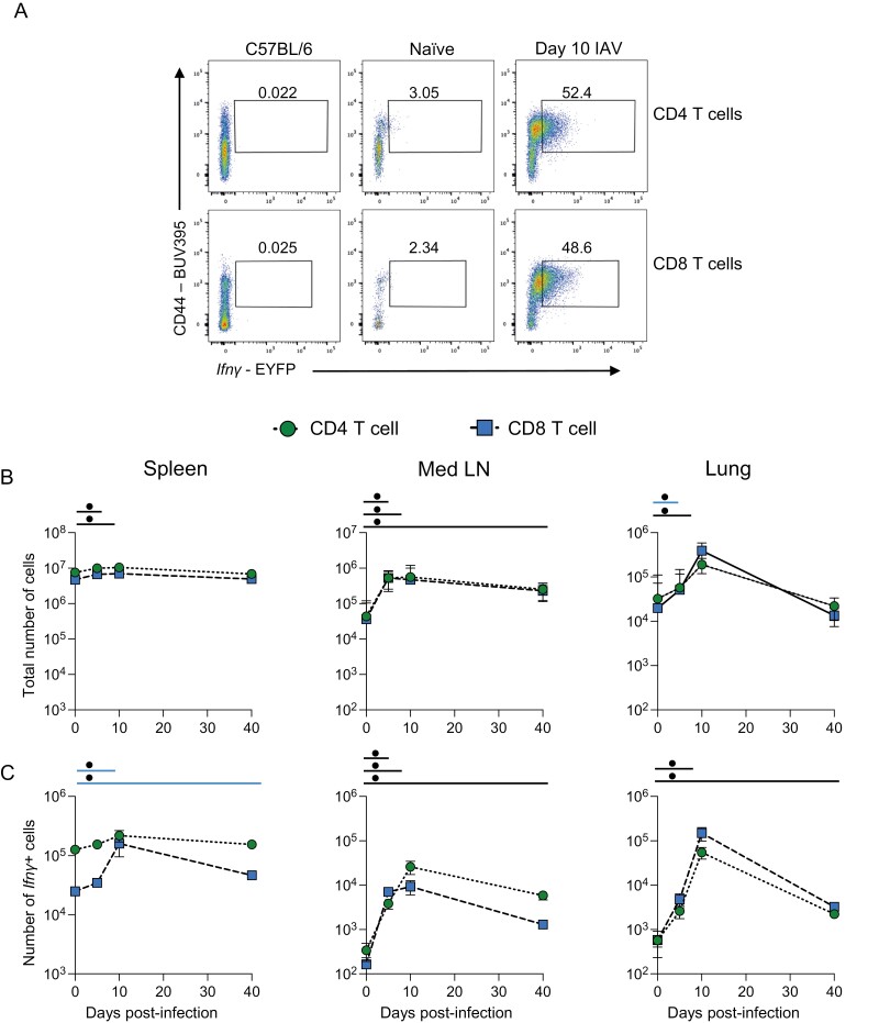
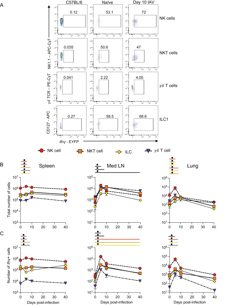
Interferon gamma (IFNγ) is a potent antiviral cytokine that can be produced by many innate and adaptive immune cells during infection. Currently, our understanding of which cells produce IFNγ and where they are located at different stages of an infection is limited. We have used reporter mice to investigate in vivo expression of Ifnγ mRNA in the lung and secondary lymphoid organs during and following influenza A virus (IAV) infection. We observed a triphasic production of Ifnγ expression. Unconventional T cells and innate lymphoid cells, particularly NK cells, were the dominant producers of early Ifnγ, while CD4 and CD8 T cells were the main producers by day 10 post-infection. Following viral clearance, some memory CD4 and CD8 T cells continued to express Ifnγ in the lungs and draining lymph node. Interestingly, Ifnγ production by lymph node natural killer (NK), NKT, and innate lymphoid type 1 cells also continued to be above naïve levels, suggesting memory-like phenotypes for these cells. Analysis of the localization of Ifnγ+ memory CD4 and CD8 T cells demonstrated that cytokine+ T cells were located near airways and in the lung parenchyma. Following a second IAV challenge, lung IAV-specific CD8 T cells rapidly increased their expression of Ifnγ while CD4 T cells in the draining lymph node increased their Ifnγ response. Together, these data suggest that Ifnγ production fluctuates based on cellular source and location, both of which could impact subsequent immune responses.


 分享
分享

 求助内容:
求助内容: 应助结果提醒方式:
 扫码关注我们
