Jordan Rosenbohm, Grayson Minnick, Bahareh Tajvidi Safa, Amir Monemian Esfahani, Xiaowei Jin, Haiwei Zhai, Nickolay V. Lavrik, Ruiguo Yang
{"title":"用双光子聚合技术制备了一种用于拉伸载荷下单细胞-细胞连接成像的多材料平台","authors":"Jordan Rosenbohm, Grayson Minnick, Bahareh Tajvidi Safa, Amir Monemian Esfahani, Xiaowei Jin, Haiwei Zhai, Nickolay V. Lavrik, Ruiguo Yang","doi":"10.1007/s10544-022-00633-z","DOIUrl":null,"url":null,"abstract":"<div><p>We previously reported a single-cell adhesion micro tensile tester (SCAμTT) fabricated from IP-S photoresin with two-photon polymerization (TPP) for investigating the mechanics of a single cell-cell junction under defined tensile loading. A major limitation of the platform is the autofluorescence of IP-S, the photoresin for TPP fabrication, which significantly increases background signal and makes fluorescent imaging of stretched cells difficult. In this study, we report the design and fabrication of a new SCAμTT platform that mitigates autofluorescence and demonstrate its capability in imaging a single cell pair as its mutual junction is stretched. By employing a two-material design using IP-S and IP-Visio, a photoresin with reduced autofluorescence, we show a significant reduction in autofluorescence of the platform. Further, by integrating apertures onto the substrate with a gold coating, the influence of autofluorescence on imaging is almost completely mitigated. With this new platform, we demonstrate the ability to image a pair of epithelial cells as they are stretched up to 250% strain, allowing us to observe junction rupture and F-actin retraction while simultaneously recording the accumulation of over 800 kPa of stress in the junction. The platform and methodology presented here can potentially enable detailed investigation of the mechanics of and mechanotransduction in cell-cell junctions and improve the design of other TPP platforms in mechanobiology applications.</p><h3>Graphical abstract</h3>\n <figure><div><div><div><picture><source><img></source></picture></div></div></div></figure>\n </div>","PeriodicalId":490,"journal":{"name":"Biomedical Microdevices","volume":"24 4","pages":""},"PeriodicalIF":3.3000,"publicationDate":"2022-10-08","publicationTypes":"Journal Article","fieldsOfStudy":null,"isOpenAccess":false,"openAccessPdf":"https://link.springer.com/content/pdf/10.1007/s10544-022-00633-z.pdf","citationCount":"2","resultStr":"{\"title\":\"A multi-material platform for imaging of single cell-cell junctions under tensile load fabricated with two-photon polymerization\",\"authors\":\"Jordan Rosenbohm, Grayson Minnick, Bahareh Tajvidi Safa, Amir Monemian Esfahani, Xiaowei Jin, Haiwei Zhai, Nickolay V. Lavrik, Ruiguo Yang\",\"doi\":\"10.1007/s10544-022-00633-z\",\"DOIUrl\":null,\"url\":null,\"abstract\":\"<div><p>We previously reported a single-cell adhesion micro tensile tester (SCAμTT) fabricated from IP-S photoresin with two-photon polymerization (TPP) for investigating the mechanics of a single cell-cell junction under defined tensile loading. A major limitation of the platform is the autofluorescence of IP-S, the photoresin for TPP fabrication, which significantly increases background signal and makes fluorescent imaging of stretched cells difficult. In this study, we report the design and fabrication of a new SCAμTT platform that mitigates autofluorescence and demonstrate its capability in imaging a single cell pair as its mutual junction is stretched. By employing a two-material design using IP-S and IP-Visio, a photoresin with reduced autofluorescence, we show a significant reduction in autofluorescence of the platform. Further, by integrating apertures onto the substrate with a gold coating, the influence of autofluorescence on imaging is almost completely mitigated. With this new platform, we demonstrate the ability to image a pair of epithelial cells as they are stretched up to 250% strain, allowing us to observe junction rupture and F-actin retraction while simultaneously recording the accumulation of over 800 kPa of stress in the junction. The platform and methodology presented here can potentially enable detailed investigation of the mechanics of and mechanotransduction in cell-cell junctions and improve the design of other TPP platforms in mechanobiology applications.</p><h3>Graphical abstract</h3>\\n <figure><div><div><div><picture><source><img></source></picture></div></div></div></figure>\\n </div>\",\"PeriodicalId\":490,\"journal\":{\"name\":\"Biomedical Microdevices\",\"volume\":\"24 4\",\"pages\":\"\"},\"PeriodicalIF\":3.3000,\"publicationDate\":\"2022-10-08\",\"publicationTypes\":\"Journal Article\",\"fieldsOfStudy\":null,\"isOpenAccess\":false,\"openAccessPdf\":\"https://link.springer.com/content/pdf/10.1007/s10544-022-00633-z.pdf\",\"citationCount\":\"2\",\"resultStr\":null,\"platform\":\"Semanticscholar\",\"paperid\":null,\"PeriodicalName\":\"Biomedical Microdevices\",\"FirstCategoryId\":\"5\",\"ListUrlMain\":\"https://link.springer.com/article/10.1007/s10544-022-00633-z\",\"RegionNum\":4,\"RegionCategory\":\"医学\",\"ArticlePicture\":[],\"TitleCN\":null,\"AbstractTextCN\":null,\"PMCID\":null,\"EPubDate\":\"\",\"PubModel\":\"\",\"JCR\":\"Q3\",\"JCRName\":\"ENGINEERING, BIOMEDICAL\",\"Score\":null,\"Total\":0}","platform":"Semanticscholar","paperid":null,"PeriodicalName":"Biomedical Microdevices","FirstCategoryId":"5","ListUrlMain":"https://link.springer.com/article/10.1007/s10544-022-00633-z","RegionNum":4,"RegionCategory":"医学","ArticlePicture":[],"TitleCN":null,"AbstractTextCN":null,"PMCID":null,"EPubDate":"","PubModel":"","JCR":"Q3","JCRName":"ENGINEERING, BIOMEDICAL","Score":null,"Total":0}
A multi-material platform for imaging of single cell-cell junctions under tensile load fabricated with two-photon polymerization
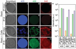
We previously reported a single-cell adhesion micro tensile tester (SCAμTT) fabricated from IP-S photoresin with two-photon polymerization (TPP) for investigating the mechanics of a single cell-cell junction under defined tensile loading. A major limitation of the platform is the autofluorescence of IP-S, the photoresin for TPP fabrication, which significantly increases background signal and makes fluorescent imaging of stretched cells difficult. In this study, we report the design and fabrication of a new SCAμTT platform that mitigates autofluorescence and demonstrate its capability in imaging a single cell pair as its mutual junction is stretched. By employing a two-material design using IP-S and IP-Visio, a photoresin with reduced autofluorescence, we show a significant reduction in autofluorescence of the platform. Further, by integrating apertures onto the substrate with a gold coating, the influence of autofluorescence on imaging is almost completely mitigated. With this new platform, we demonstrate the ability to image a pair of epithelial cells as they are stretched up to 250% strain, allowing us to observe junction rupture and F-actin retraction while simultaneously recording the accumulation of over 800 kPa of stress in the junction. The platform and methodology presented here can potentially enable detailed investigation of the mechanics of and mechanotransduction in cell-cell junctions and improve the design of other TPP platforms in mechanobiology applications.
期刊介绍:
Biomedical Microdevices: BioMEMS and Biomedical Nanotechnology is an interdisciplinary periodical devoted to all aspects of research in the medical diagnostic and therapeutic applications of Micro-Electro-Mechanical Systems (BioMEMS) and nanotechnology for medicine and biology.
General subjects of interest include the design, characterization, testing, modeling and clinical validation of microfabricated systems, and their integration on-chip and in larger functional units. The specific interests of the Journal include systems for neural stimulation and recording, bioseparation technologies such as nanofilters and electrophoretic equipment, miniaturized analytic and DNA identification systems, biosensors, and micro/nanotechnologies for cell and tissue research, tissue engineering, cell transplantation, and the controlled release of drugs and biological molecules.
Contributions reporting on fundamental and applied investigations of the material science, biochemistry, and physics of biomedical microdevices and nanotechnology are encouraged. A non-exhaustive list of fields of interest includes: nanoparticle synthesis, characterization, and validation of therapeutic or imaging efficacy in animal models; biocompatibility; biochemical modification of microfabricated devices, with reference to non-specific protein adsorption, and the active immobilization and patterning of proteins on micro/nanofabricated surfaces; the dynamics of fluids in micro-and-nano-fabricated channels; the electromechanical and structural response of micro/nanofabricated systems; the interactions of microdevices with cells and tissues, including biocompatibility and biodegradation studies; variations in the characteristics of the systems as a function of the micro/nanofabrication parameters.


 分享
分享
 求助内容:
求助内容: 应助结果提醒方式:
 扫码关注我们
