Magnus Nord, Per Erik Vullum, Ian MacLaren, Thomas Tybell, Randi Holmestad
{"title":"Atomap:一个使用二维高斯拟合自动分析原子分辨率图像的新软件工具","authors":"Magnus Nord, Per Erik Vullum, Ian MacLaren, Thomas Tybell, Randi Holmestad","doi":"10.1186/s40679-017-0042-5","DOIUrl":null,"url":null,"abstract":"<p>Scanning transmission electron microscopy (STEM) data with atomic resolution can contain a large amount of information about the structure of a crystalline material. Often, this information is hard to extract, due to the large number of atomic columns and large differences in intensity from sublattices consisting of different elements. In this work, we present a free and open source software tool for analysing both the position and shapes of atomic columns in STEM-images, using 2-D elliptical Gaussian distributions. The software is tested on variants of the perovskite oxide structure. By first fitting the most intense atomic columns and then subtracting them, information on all the projected sublattices can be obtained. From this, we can extract changes in the lattice parameters and shape of A-cation columns from annular dark field images of perovskite oxide heterostructures. Using annular bright field images, shifts in oxygen column positions are also quantified in the same heterostructure. The precision of determining the position of atomic columns is compared between STEM data acquired using standard acquisition, and STEM-images obtained as an image stack averaged after using non-rigid registration.</p>","PeriodicalId":460,"journal":{"name":"Advanced Structural and Chemical Imaging","volume":"3 1","pages":""},"PeriodicalIF":3.5600,"publicationDate":"2017-02-13","publicationTypes":"Journal Article","fieldsOfStudy":null,"isOpenAccess":false,"openAccessPdf":"https://sci-hub-pdf.com/10.1186/s40679-017-0042-5","citationCount":"154","resultStr":"{\"title\":\"Atomap: a new software tool for the automated analysis of atomic resolution images using two-dimensional Gaussian fitting\",\"authors\":\"Magnus Nord, Per Erik Vullum, Ian MacLaren, Thomas Tybell, Randi Holmestad\",\"doi\":\"10.1186/s40679-017-0042-5\",\"DOIUrl\":null,\"url\":null,\"abstract\":\"<p>Scanning transmission electron microscopy (STEM) data with atomic resolution can contain a large amount of information about the structure of a crystalline material. Often, this information is hard to extract, due to the large number of atomic columns and large differences in intensity from sublattices consisting of different elements. In this work, we present a free and open source software tool for analysing both the position and shapes of atomic columns in STEM-images, using 2-D elliptical Gaussian distributions. The software is tested on variants of the perovskite oxide structure. By first fitting the most intense atomic columns and then subtracting them, information on all the projected sublattices can be obtained. From this, we can extract changes in the lattice parameters and shape of A-cation columns from annular dark field images of perovskite oxide heterostructures. Using annular bright field images, shifts in oxygen column positions are also quantified in the same heterostructure. The precision of determining the position of atomic columns is compared between STEM data acquired using standard acquisition, and STEM-images obtained as an image stack averaged after using non-rigid registration.</p>\",\"PeriodicalId\":460,\"journal\":{\"name\":\"Advanced Structural and Chemical Imaging\",\"volume\":\"3 1\",\"pages\":\"\"},\"PeriodicalIF\":3.5600,\"publicationDate\":\"2017-02-13\",\"publicationTypes\":\"Journal Article\",\"fieldsOfStudy\":null,\"isOpenAccess\":false,\"openAccessPdf\":\"https://sci-hub-pdf.com/10.1186/s40679-017-0042-5\",\"citationCount\":\"154\",\"resultStr\":null,\"platform\":\"Semanticscholar\",\"paperid\":null,\"PeriodicalName\":\"Advanced Structural and Chemical Imaging\",\"FirstCategoryId\":\"1085\",\"ListUrlMain\":\"https://link.springer.com/article/10.1186/s40679-017-0042-5\",\"RegionNum\":0,\"RegionCategory\":null,\"ArticlePicture\":[],\"TitleCN\":null,\"AbstractTextCN\":null,\"PMCID\":null,\"EPubDate\":\"\",\"PubModel\":\"\",\"JCR\":\"Q1\",\"JCRName\":\"Medicine\",\"Score\":null,\"Total\":0}","platform":"Semanticscholar","paperid":null,"PeriodicalName":"Advanced Structural and Chemical Imaging","FirstCategoryId":"1085","ListUrlMain":"https://link.springer.com/article/10.1186/s40679-017-0042-5","RegionNum":0,"RegionCategory":null,"ArticlePicture":[],"TitleCN":null,"AbstractTextCN":null,"PMCID":null,"EPubDate":"","PubModel":"","JCR":"Q1","JCRName":"Medicine","Score":null,"Total":0}
Atomap: a new software tool for the automated analysis of atomic resolution images using two-dimensional Gaussian fitting
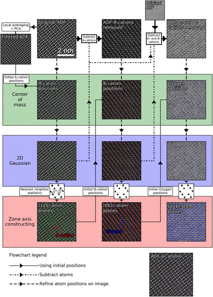
Scanning transmission electron microscopy (STEM) data with atomic resolution can contain a large amount of information about the structure of a crystalline material. Often, this information is hard to extract, due to the large number of atomic columns and large differences in intensity from sublattices consisting of different elements. In this work, we present a free and open source software tool for analysing both the position and shapes of atomic columns in STEM-images, using 2-D elliptical Gaussian distributions. The software is tested on variants of the perovskite oxide structure. By first fitting the most intense atomic columns and then subtracting them, information on all the projected sublattices can be obtained. From this, we can extract changes in the lattice parameters and shape of A-cation columns from annular dark field images of perovskite oxide heterostructures. Using annular bright field images, shifts in oxygen column positions are also quantified in the same heterostructure. The precision of determining the position of atomic columns is compared between STEM data acquired using standard acquisition, and STEM-images obtained as an image stack averaged after using non-rigid registration.


 分享
分享
 求助内容:
求助内容: 应助结果提醒方式:
 扫码关注我们
