Vishal Singh, Jaladhar Neelavalli, Suhail P Parvaze, Mamta Gupta, Radha K Verma, Avnish K Seth, Lakshay Mehta, Rakesh Kumar Gupta
{"title":"与传统MRI相比,磁化率加权成像在检测胆总管结石中的相对贡献。","authors":"Vishal Singh, Jaladhar Neelavalli, Suhail P Parvaze, Mamta Gupta, Radha K Verma, Avnish K Seth, Lakshay Mehta, Rakesh Kumar Gupta","doi":"10.5152/dir.2022.20713","DOIUrl":null,"url":null,"abstract":"<p><p>PURPOSE We aimed to evaluate the relative contribution of susceptibility weighted imaging (SWI) in the detection of common bile-duct (CBD) stones in comparison to the conventional MRI protocol containing magnetic resonance cholangiopancreatography (MRCP), balanced turbo field echo (BTFE), and T2-weighted spin-echo imaging techniques. METHODS MRI data containing MRCP, BTFE, T2-weighted imaging, and abdominal SWI were independently evaluated by 2 sets of experienced radiologists in 44 patients with confirmed CBD stones. Endoscopic retrograde cholangiopancreatography, and endoscopic ultrasound where available, was used as the reference gold standard. Evaluation was performed for the visualization of CBD stones in each of the MRI techniques. Relative contribution of SWI was classified into one of four categories for each case: (1) no contribution to CBD stone visualization; (2) same as conventional techniques; (3) improved diagnostic confidence; and (4) critical for diagnosis. Stone size was also assessed. RESULTS Inter-rater agreement coefficient for CBD stone visualization was found to be \"good\" in MRCP (0.77), \"very good\" in SWI (0.94) and BTFE (0.84), and moderate in T2-weighted imaging (0.54). CBD stones were visualized with SWI in 86.4% and 82%, with MRCP in 70.5% and 70.5% cases, with BTFE in 73% and 61.4% cases, with T2-weighted imaging in 45.5% and 52.3% cases by reviewers 1 and 2, respectively. SWI did not contribute to CBD stone visualization in 2.3% (1/44); was the same as conventional techniques in 31.8% (14/44) cases; improved diagnostic confidence in 34.1%; and was critical for diagnosis in 20.5% cases. CONCLUSION SWI has the potential to serve as a strong adjunct to conventional MRI protocols used for CBD stone evaluation with very small scan-time penalty.</p>","PeriodicalId":50582,"journal":{"name":"Diagnostic and Interventional Radiology","volume":"28 2 1","pages":"131-137"},"PeriodicalIF":1.7000,"publicationDate":"2022-03-01","publicationTypes":"Journal Article","fieldsOfStudy":null,"isOpenAccess":false,"openAccessPdf":"https://www.ncbi.nlm.nih.gov/pmc/articles/PMC12278929/pdf/","citationCount":"0","resultStr":"{\"title\":\"Relative contribution of susceptibility weighted imaging, compared to conventional MRI, in the detection of common bile-duct calculi.\",\"authors\":\"Vishal Singh, Jaladhar Neelavalli, Suhail P Parvaze, Mamta Gupta, Radha K Verma, Avnish K Seth, Lakshay Mehta, Rakesh Kumar Gupta\",\"doi\":\"10.5152/dir.2022.20713\",\"DOIUrl\":null,\"url\":null,\"abstract\":\"<p><p>PURPOSE We aimed to evaluate the relative contribution of susceptibility weighted imaging (SWI) in the detection of common bile-duct (CBD) stones in comparison to the conventional MRI protocol containing magnetic resonance cholangiopancreatography (MRCP), balanced turbo field echo (BTFE), and T2-weighted spin-echo imaging techniques. METHODS MRI data containing MRCP, BTFE, T2-weighted imaging, and abdominal SWI were independently evaluated by 2 sets of experienced radiologists in 44 patients with confirmed CBD stones. Endoscopic retrograde cholangiopancreatography, and endoscopic ultrasound where available, was used as the reference gold standard. Evaluation was performed for the visualization of CBD stones in each of the MRI techniques. Relative contribution of SWI was classified into one of four categories for each case: (1) no contribution to CBD stone visualization; (2) same as conventional techniques; (3) improved diagnostic confidence; and (4) critical for diagnosis. Stone size was also assessed. RESULTS Inter-rater agreement coefficient for CBD stone visualization was found to be \\\"good\\\" in MRCP (0.77), \\\"very good\\\" in SWI (0.94) and BTFE (0.84), and moderate in T2-weighted imaging (0.54). CBD stones were visualized with SWI in 86.4% and 82%, with MRCP in 70.5% and 70.5% cases, with BTFE in 73% and 61.4% cases, with T2-weighted imaging in 45.5% and 52.3% cases by reviewers 1 and 2, respectively. SWI did not contribute to CBD stone visualization in 2.3% (1/44); was the same as conventional techniques in 31.8% (14/44) cases; improved diagnostic confidence in 34.1%; and was critical for diagnosis in 20.5% cases. CONCLUSION SWI has the potential to serve as a strong adjunct to conventional MRI protocols used for CBD stone evaluation with very small scan-time penalty.</p>\",\"PeriodicalId\":50582,\"journal\":{\"name\":\"Diagnostic and Interventional Radiology\",\"volume\":\"28 2 1\",\"pages\":\"131-137\"},\"PeriodicalIF\":1.7000,\"publicationDate\":\"2022-03-01\",\"publicationTypes\":\"Journal Article\",\"fieldsOfStudy\":null,\"isOpenAccess\":false,\"openAccessPdf\":\"https://www.ncbi.nlm.nih.gov/pmc/articles/PMC12278929/pdf/\",\"citationCount\":\"0\",\"resultStr\":null,\"platform\":\"Semanticscholar\",\"paperid\":null,\"PeriodicalName\":\"Diagnostic and Interventional Radiology\",\"FirstCategoryId\":\"3\",\"ListUrlMain\":\"https://doi.org/10.5152/dir.2022.20713\",\"RegionNum\":4,\"RegionCategory\":\"医学\",\"ArticlePicture\":[],\"TitleCN\":null,\"AbstractTextCN\":null,\"PMCID\":null,\"EPubDate\":\"\",\"PubModel\":\"\",\"JCR\":\"Q2\",\"JCRName\":\"Medicine\",\"Score\":null,\"Total\":0}","platform":"Semanticscholar","paperid":null,"PeriodicalName":"Diagnostic and Interventional Radiology","FirstCategoryId":"3","ListUrlMain":"https://doi.org/10.5152/dir.2022.20713","RegionNum":4,"RegionCategory":"医学","ArticlePicture":[],"TitleCN":null,"AbstractTextCN":null,"PMCID":null,"EPubDate":"","PubModel":"","JCR":"Q2","JCRName":"Medicine","Score":null,"Total":0}
Relative contribution of susceptibility weighted imaging, compared to conventional MRI, in the detection of common bile-duct calculi.
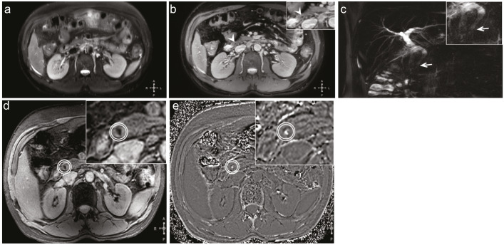
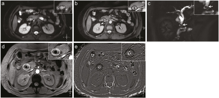
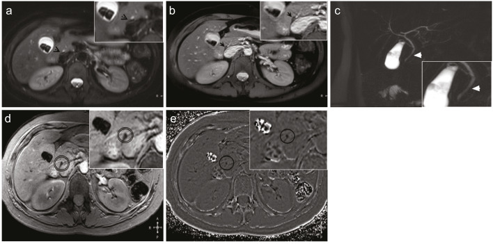
PURPOSE We aimed to evaluate the relative contribution of susceptibility weighted imaging (SWI) in the detection of common bile-duct (CBD) stones in comparison to the conventional MRI protocol containing magnetic resonance cholangiopancreatography (MRCP), balanced turbo field echo (BTFE), and T2-weighted spin-echo imaging techniques. METHODS MRI data containing MRCP, BTFE, T2-weighted imaging, and abdominal SWI were independently evaluated by 2 sets of experienced radiologists in 44 patients with confirmed CBD stones. Endoscopic retrograde cholangiopancreatography, and endoscopic ultrasound where available, was used as the reference gold standard. Evaluation was performed for the visualization of CBD stones in each of the MRI techniques. Relative contribution of SWI was classified into one of four categories for each case: (1) no contribution to CBD stone visualization; (2) same as conventional techniques; (3) improved diagnostic confidence; and (4) critical for diagnosis. Stone size was also assessed. RESULTS Inter-rater agreement coefficient for CBD stone visualization was found to be "good" in MRCP (0.77), "very good" in SWI (0.94) and BTFE (0.84), and moderate in T2-weighted imaging (0.54). CBD stones were visualized with SWI in 86.4% and 82%, with MRCP in 70.5% and 70.5% cases, with BTFE in 73% and 61.4% cases, with T2-weighted imaging in 45.5% and 52.3% cases by reviewers 1 and 2, respectively. SWI did not contribute to CBD stone visualization in 2.3% (1/44); was the same as conventional techniques in 31.8% (14/44) cases; improved diagnostic confidence in 34.1%; and was critical for diagnosis in 20.5% cases. CONCLUSION SWI has the potential to serve as a strong adjunct to conventional MRI protocols used for CBD stone evaluation with very small scan-time penalty.
期刊介绍:
Diagnostic and Interventional Radiology (Diagn Interv Radiol) is the open access, online-only official publication of Turkish Society of Radiology. It is published bimonthly and the journal’s publication language is English.
The journal is a medium for original articles, reviews, pictorial essays, technical notes related to all fields of diagnostic and interventional radiology.


 分享
分享
 求助内容:
求助内容: 应助结果提醒方式:
 扫码关注我们
