Lu Lv, En-Hai Cui, Bin Wang, Li-Qin Li, Feng Hua, Hua-Dong Lu, Na Chen, Wen-Yan Chen
{"title":"多组学显示人脐带间充质干细胞通过肺肠轴改善急性肺损伤。","authors":"Lu Lv, En-Hai Cui, Bin Wang, Li-Qin Li, Feng Hua, Hua-Dong Lu, Na Chen, Wen-Yan Chen","doi":"10.4252/wjsc.v15.i9.908","DOIUrl":null,"url":null,"abstract":"<p><strong>Background: </strong>Acute lung injury (ALI) and its final severe stage, acute respiratory distress syndrome, are associated with high morbidity and mortality rates in patients due to the lack of effective specific treatments. Gut microbiota homeostasis, including that in ALI, is important for human health. Evidence suggests that the gut microbiota improves lung injury through the lung-gut axis. Human umbilical cord mesenchymal cells (HUC-MSCs) have attractive prospects for ALI treatment. This study hypothesized that HUC-MSCs improve ALI <i>via</i> the lung-gut microflora.</p><p><strong>Aim: </strong>To explore the effects of HUC-MSCs on lipopolysaccharide (LPS)-induced ALI in mice and the involvement of the lung-gut axis in this process.</p><p><strong>Methods: </strong>C57BL/6 mice were randomly divided into four groups (18 rats per group): Sham, sham + HUC-MSCs, LPS, and LPS + HUC-MSCs. ALI was induced in mice by intraperitoneal injections of LPS (10 mg/kg). After 6 h, mice were intervened with 0.5 mL phosphate buffered saline (PBS) containing 1 × 10<sup>6</sup> HUC-MSCs by intraperitoneal injections. For the negative control, 100 mL 0.9% NaCl and 0.5 mL PBS were used. Bronchoalveolar lavage fluid (BALF) was obtained from anesthetized mice, and their blood, lungs, ileum, and feces were obtained by an aseptic technique following CO<sub>2</sub> euthanasia. Wright's staining, enzyme-linked immunosorbent assay, hematoxylin-eosin staining, Evans blue dye leakage assay, immunohistochemistry, fluorescence <i>in situ</i> hybridization, western blot, 16S rDNA sequencing, and non-targeted metabolomics were used to observe the effect of HUC-MSCs on ALI mice, and the involvement of the lung-gut axis in this process was explored. One-way analysis of variance with post-hoc Tukey's test, independent-sample Student's <i>t</i>-test, Wilcoxon rank-sum test, and Pearson correlation analysis were used for statistical analyses.</p><p><strong>Results: </strong>HUC-MSCs were observed to improve pulmonary edema and lung and ileal injury, and decrease mononuclear cell and neutrophil counts, protein concentrations in BALF and inflammatory cytokine levels in the serum, lung, and ileum of ALI mice. Especially, HUC-MSCs decreased Evans blue concentration and Toll-like receptor 4, myeloid differentiation factor 88, p-nuclear factor kappa-B (NF-κB)/NF-κB, and p-inhibitor α of NF-κB (p-IκBα)/IκBα expression levels in the lung, and raised the pulmonary vascular endothelial-cadherin, zonula occludens-1 (ZO-1), and occludin levels and ileal ZO-1, claudin-1, and occludin expression levels. HUC-MSCs improved gut and BALF microbial homeostases. The number of pathogenic bacteria decreased in the BALF of ALI mice treated with HUC-MSCs. Concurrently, the abundances of <i>Oscillospira</i> and <i>Coprococcus</i> in the feces of HUS-MSC-treated ALI mice were significantly increased. In addition, <i>Lactobacillus</i>, <i>Bacteroides</i>, and <i>unidentified_Rikenellaceae</i> genera appeared in both feces and BALF. Moreover, this study performed metabolomic analysis on the lung tissue and identified five upregulated metabolites and 11 downregulated metabolites in the LPS + MSC group compared to the LPS group, which were related to the purine metabolism and the taste transduction signaling pathways. Therefore, an intrinsic link between lung metabolite levels and BALF flora homeostasis was established.</p><p><strong>Conclusion: </strong>This study suggests that HUM-MSCs attenuate ALI by redefining the gut and lung microbiota.</p>","PeriodicalId":23775,"journal":{"name":"World journal of stem cells","volume":"15 9","pages":"908-930"},"PeriodicalIF":3.6000,"publicationDate":"2023-09-26","publicationTypes":"Journal Article","fieldsOfStudy":null,"isOpenAccess":false,"openAccessPdf":"https://www.ncbi.nlm.nih.gov/pmc/articles/PMC10600741/pdf/","citationCount":"0","resultStr":"{\"title\":\"Multiomics reveal human umbilical cord mesenchymal stem cells improving acute lung injury <i>via</i> the lung-gut axis.\",\"authors\":\"Lu Lv, En-Hai Cui, Bin Wang, Li-Qin Li, Feng Hua, Hua-Dong Lu, Na Chen, Wen-Yan Chen\",\"doi\":\"10.4252/wjsc.v15.i9.908\",\"DOIUrl\":null,\"url\":null,\"abstract\":\"<p><strong>Background: </strong>Acute lung injury (ALI) and its final severe stage, acute respiratory distress syndrome, are associated with high morbidity and mortality rates in patients due to the lack of effective specific treatments. Gut microbiota homeostasis, including that in ALI, is important for human health. Evidence suggests that the gut microbiota improves lung injury through the lung-gut axis. Human umbilical cord mesenchymal cells (HUC-MSCs) have attractive prospects for ALI treatment. This study hypothesized that HUC-MSCs improve ALI <i>via</i> the lung-gut microflora.</p><p><strong>Aim: </strong>To explore the effects of HUC-MSCs on lipopolysaccharide (LPS)-induced ALI in mice and the involvement of the lung-gut axis in this process.</p><p><strong>Methods: </strong>C57BL/6 mice were randomly divided into four groups (18 rats per group): Sham, sham + HUC-MSCs, LPS, and LPS + HUC-MSCs. ALI was induced in mice by intraperitoneal injections of LPS (10 mg/kg). After 6 h, mice were intervened with 0.5 mL phosphate buffered saline (PBS) containing 1 × 10<sup>6</sup> HUC-MSCs by intraperitoneal injections. For the negative control, 100 mL 0.9% NaCl and 0.5 mL PBS were used. Bronchoalveolar lavage fluid (BALF) was obtained from anesthetized mice, and their blood, lungs, ileum, and feces were obtained by an aseptic technique following CO<sub>2</sub> euthanasia. Wright's staining, enzyme-linked immunosorbent assay, hematoxylin-eosin staining, Evans blue dye leakage assay, immunohistochemistry, fluorescence <i>in situ</i> hybridization, western blot, 16S rDNA sequencing, and non-targeted metabolomics were used to observe the effect of HUC-MSCs on ALI mice, and the involvement of the lung-gut axis in this process was explored. One-way analysis of variance with post-hoc Tukey's test, independent-sample Student's <i>t</i>-test, Wilcoxon rank-sum test, and Pearson correlation analysis were used for statistical analyses.</p><p><strong>Results: </strong>HUC-MSCs were observed to improve pulmonary edema and lung and ileal injury, and decrease mononuclear cell and neutrophil counts, protein concentrations in BALF and inflammatory cytokine levels in the serum, lung, and ileum of ALI mice. Especially, HUC-MSCs decreased Evans blue concentration and Toll-like receptor 4, myeloid differentiation factor 88, p-nuclear factor kappa-B (NF-κB)/NF-κB, and p-inhibitor α of NF-κB (p-IκBα)/IκBα expression levels in the lung, and raised the pulmonary vascular endothelial-cadherin, zonula occludens-1 (ZO-1), and occludin levels and ileal ZO-1, claudin-1, and occludin expression levels. HUC-MSCs improved gut and BALF microbial homeostases. The number of pathogenic bacteria decreased in the BALF of ALI mice treated with HUC-MSCs. Concurrently, the abundances of <i>Oscillospira</i> and <i>Coprococcus</i> in the feces of HUS-MSC-treated ALI mice were significantly increased. In addition, <i>Lactobacillus</i>, <i>Bacteroides</i>, and <i>unidentified_Rikenellaceae</i> genera appeared in both feces and BALF. Moreover, this study performed metabolomic analysis on the lung tissue and identified five upregulated metabolites and 11 downregulated metabolites in the LPS + MSC group compared to the LPS group, which were related to the purine metabolism and the taste transduction signaling pathways. Therefore, an intrinsic link between lung metabolite levels and BALF flora homeostasis was established.</p><p><strong>Conclusion: </strong>This study suggests that HUM-MSCs attenuate ALI by redefining the gut and lung microbiota.</p>\",\"PeriodicalId\":23775,\"journal\":{\"name\":\"World journal of stem cells\",\"volume\":\"15 9\",\"pages\":\"908-930\"},\"PeriodicalIF\":3.6000,\"publicationDate\":\"2023-09-26\",\"publicationTypes\":\"Journal Article\",\"fieldsOfStudy\":null,\"isOpenAccess\":false,\"openAccessPdf\":\"https://www.ncbi.nlm.nih.gov/pmc/articles/PMC10600741/pdf/\",\"citationCount\":\"0\",\"resultStr\":null,\"platform\":\"Semanticscholar\",\"paperid\":null,\"PeriodicalName\":\"World journal of stem cells\",\"FirstCategoryId\":\"3\",\"ListUrlMain\":\"https://doi.org/10.4252/wjsc.v15.i9.908\",\"RegionNum\":3,\"RegionCategory\":\"医学\",\"ArticlePicture\":[],\"TitleCN\":null,\"AbstractTextCN\":null,\"PMCID\":null,\"EPubDate\":\"\",\"PubModel\":\"\",\"JCR\":\"Q3\",\"JCRName\":\"CELL & TISSUE ENGINEERING\",\"Score\":null,\"Total\":0}","platform":"Semanticscholar","paperid":null,"PeriodicalName":"World journal of stem cells","FirstCategoryId":"3","ListUrlMain":"https://doi.org/10.4252/wjsc.v15.i9.908","RegionNum":3,"RegionCategory":"医学","ArticlePicture":[],"TitleCN":null,"AbstractTextCN":null,"PMCID":null,"EPubDate":"","PubModel":"","JCR":"Q3","JCRName":"CELL & TISSUE ENGINEERING","Score":null,"Total":0}
引用次数: 0
摘要
背景:由于缺乏有效的特异性治疗,急性肺损伤(ALI)及其最后的严重阶段急性呼吸窘迫综合征与患者的高发病率和死亡率有关。肠道微生物群的稳态,包括ALI的稳态,对人类健康很重要。有证据表明,肠道微生物群通过肺-肠轴改善肺损伤。人脐带间充质细胞(HUC-MSCs)在治疗急性肺损伤方面具有很好的前景。本研究假设HUC-MSCs通过肺肠道菌群改善ALI。目的:探讨HUC-MSCs对脂多糖(LPS)诱导的小鼠急性肺损伤(ALI)的影响及其与肺肠轴的关系。方法:将C57BL/6小鼠随机分为四组(每组18只):Sham、Sham+HUC-MSCs、LPS和LPS+HUC-MSC。通过腹膜内注射LPS(10mg/kg)在小鼠中诱导ALI。6小时后,通过腹膜内注射用含有1×106个HUC MSC的0.5mL磷酸盐缓冲盐水(PBS)干预小鼠。对于阴性对照,使用100 mL 0.9%NaCl和0.5 mL PBS。从麻醉小鼠获得支气管肺泡灌洗液(BALF),并在CO2安乐死后通过无菌技术获得它们的血液、肺、回肠和粪便。采用Wright染色、酶联免疫吸附试验、苏木精-伊红染色、Evans蓝染料渗漏试验、免疫组织化学、荧光原位杂交、蛋白质印迹、16S rDNA测序和非靶向代谢组学等方法观察HUC-MSCs对ALI小鼠的影响,并探讨肺肠轴在这一过程中的作用。统计学分析采用单因素方差分析,包括事后Tukey检验、独立样本Student t检验、Wilcoxon秩和检验和Pearson相关分析。结果:HUC-MSCs可改善ALI小鼠的肺水肿、肺损伤和回肠损伤,并降低血清、肺和回肠中的单核细胞和中性粒细胞计数、BALF中的蛋白质浓度以及炎症细胞因子水平。特别是,HUC-MSCs降低了肺中Evans蓝浓度和Toll样受体4、骨髓分化因子88、p-核因子κB(NF-κB)/NF-κB和p-κB抑制剂α(p-IκBα)/IκBα的表达水平,并升高了肺血管内皮钙粘蛋白、闭塞带-1(ZO-1)和闭塞素水平以及回肠ZO-1、claudin-1和闭塞素表达水平。HUC-MSCs改善了肠道和BALF微生物的稳态。HUC-MSCs处理的ALI小鼠BALF中致病菌数量减少。同时,HUS-MSC处理的ALI小鼠粪便中的示波螺旋菌和球菌的丰度显著增加。此外,在粪便和BALF中都出现了乳酸杆菌属、拟杆菌属和未识别的里氏杆菌科属。此外,本研究对肺组织进行了代谢组学分析,发现与LPS组相比,LPS+MSC组有5种上调代谢产物和11种下调代谢产物,这与嘌呤代谢和味觉转导信号通路有关。因此,建立了肺代谢产物水平和BALF菌群稳态之间的内在联系。结论:本研究表明HUM-MSCs通过重新定义肠道和肺部微生物群来减轻ALI。
Multiomics reveal human umbilical cord mesenchymal stem cells improving acute lung injury via the lung-gut axis.
Background: Acute lung injury (ALI) and its final severe stage, acute respiratory distress syndrome, are associated with high morbidity and mortality rates in patients due to the lack of effective specific treatments. Gut microbiota homeostasis, including that in ALI, is important for human health. Evidence suggests that the gut microbiota improves lung injury through the lung-gut axis. Human umbilical cord mesenchymal cells (HUC-MSCs) have attractive prospects for ALI treatment. This study hypothesized that HUC-MSCs improve ALI via the lung-gut microflora.
Aim: To explore the effects of HUC-MSCs on lipopolysaccharide (LPS)-induced ALI in mice and the involvement of the lung-gut axis in this process.
Methods: C57BL/6 mice were randomly divided into four groups (18 rats per group): Sham, sham + HUC-MSCs, LPS, and LPS + HUC-MSCs. ALI was induced in mice by intraperitoneal injections of LPS (10 mg/kg). After 6 h, mice were intervened with 0.5 mL phosphate buffered saline (PBS) containing 1 × 10⁶ HUC-MSCs by intraperitoneal injections. For the negative control, 100 mL 0.9% NaCl and 0.5 mL PBS were used. Bronchoalveolar lavage fluid (BALF) was obtained from anesthetized mice, and their blood, lungs, ileum, and feces were obtained by an aseptic technique following CO2 euthanasia. Wright's staining, enzyme-linked immunosorbent assay, hematoxylin-eosin staining, Evans blue dye leakage assay, immunohistochemistry, fluorescence in situ hybridization, western blot, 16S rDNA sequencing, and non-targeted metabolomics were used to observe the effect of HUC-MSCs on ALI mice, and the involvement of the lung-gut axis in this process was explored. One-way analysis of variance with post-hoc Tukey's test, independent-sample Student's t-test, Wilcoxon rank-sum test, and Pearson correlation analysis were used for statistical analyses.
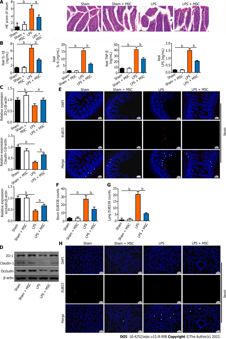
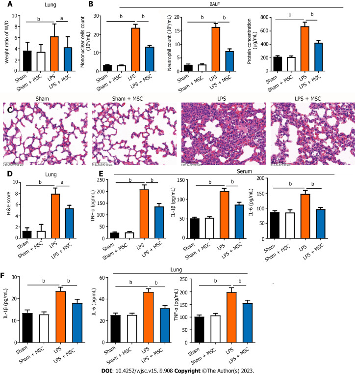
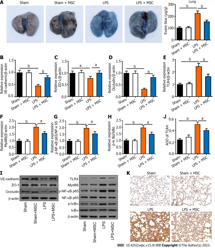
Results: HUC-MSCs were observed to improve pulmonary edema and lung and ileal injury, and decrease mononuclear cell and neutrophil counts, protein concentrations in BALF and inflammatory cytokine levels in the serum, lung, and ileum of ALI mice. Especially, HUC-MSCs decreased Evans blue concentration and Toll-like receptor 4, myeloid differentiation factor 88, p-nuclear factor kappa-B (NF-κB)/NF-κB, and p-inhibitor α of NF-κB (p-IκBα)/IκBα expression levels in the lung, and raised the pulmonary vascular endothelial-cadherin, zonula occludens-1 (ZO-1), and occludin levels and ileal ZO-1, claudin-1, and occludin expression levels. HUC-MSCs improved gut and BALF microbial homeostases. The number of pathogenic bacteria decreased in the BALF of ALI mice treated with HUC-MSCs. Concurrently, the abundances of Oscillospira and Coprococcus in the feces of HUS-MSC-treated ALI mice were significantly increased. In addition, Lactobacillus, Bacteroides, and unidentified_Rikenellaceae genera appeared in both feces and BALF. Moreover, this study performed metabolomic analysis on the lung tissue and identified five upregulated metabolites and 11 downregulated metabolites in the LPS + MSC group compared to the LPS group, which were related to the purine metabolism and the taste transduction signaling pathways. Therefore, an intrinsic link between lung metabolite levels and BALF flora homeostasis was established.
Conclusion: This study suggests that HUM-MSCs attenuate ALI by redefining the gut and lung microbiota.
期刊介绍:
The World Journal of Stem Cells (WJSC) is a leading academic journal devoted to reporting the latest, cutting-edge research progress and findings of basic research and clinical practice in the field of stem cells. It was launched on December 31, 2009 and is published monthly (12 issues annually) by BPG, the world''s leading professional clinical medical journal publishing company.


 分享
分享
 求助内容:
求助内容: 应助结果提醒方式:
 扫码关注我们
