{"title":"锥形束计算机断层扫描下颌舌骨与第三磨牙危险因素的定量和定性相关性。","authors":"Mehuli Halder, Yogesh Chhaparwal, Vathsala Patil, Komal Smriti, Shubha Chhaparwal, Kalyana C Pentapati","doi":"10.2147/CCIDE.S428908","DOIUrl":null,"url":null,"abstract":"<p><strong>Background: </strong>Lingual plate thickness, density, and proximity to the tooth are linked as risk factors for various complications associated with third molar extraction. The present study aimed to assess the lingual plate thickness, and density in the mandibular third molar region using cone beam computed tomography and to estimate its correlation with type and level of impaction, number of roots, age, and gender as the risk factors.</p><p><strong>Methods: </strong>This was a retrospective study on CBCT images of 648 mandibular third molars. The lingual plate thickness at three different root levels - cervical, mid-root, and apex along with the position of the tooth, number of roots, density of lingual plate, age, and gender were evaluated. The measurements were done on Invivo 5-Anatomage software. Statistical comparison of the categorical variables was done by Chi-square test, and Fisher's exact test, and univariate and multivariate analysis were done using binomial logistic regression.</p><p><strong>Results: </strong>Lingual plate thickness of the third molars at the cervical, mid root, and apex were 1.28 mm, 1.42 mm and .01 mm (mean). A significantly higher proportion of subjects with thin lingual plates at mid-root (p-value=0.01) and apex (p-value=0.05) were in the 21-30 age group. Lingual bone density was significantly associated with the thickness of the lingual plate at the mid-root. A significantly higher proportion of thinner lingual plates at the mid-root level were associated with mesioangularly placed third molars (p-value=0.002).</p><p><strong>Conclusion: </strong>Our study presented that lingual plate thickness has a strong association with age, angulation, and the number of roots. Knowledge about these risk factors is imperative during the management of third molar impactions.</p>","PeriodicalId":10445,"journal":{"name":"Clinical, Cosmetic and Investigational Dentistry","volume":"15 ","pages":"267-277"},"PeriodicalIF":1.8000,"publicationDate":"2023-10-30","publicationTypes":"Journal Article","fieldsOfStudy":null,"isOpenAccess":false,"openAccessPdf":"https://www.ncbi.nlm.nih.gov/pmc/articles/PMC10624182/pdf/","citationCount":"0","resultStr":"{\"title\":\"Quantitative and Qualitative Correlation of Mandibular Lingual Bone with Risk Factors for Third Molar Using Cone Beam Computed Tomography.\",\"authors\":\"Mehuli Halder, Yogesh Chhaparwal, Vathsala Patil, Komal Smriti, Shubha Chhaparwal, Kalyana C Pentapati\",\"doi\":\"10.2147/CCIDE.S428908\",\"DOIUrl\":null,\"url\":null,\"abstract\":\"<p><strong>Background: </strong>Lingual plate thickness, density, and proximity to the tooth are linked as risk factors for various complications associated with third molar extraction. The present study aimed to assess the lingual plate thickness, and density in the mandibular third molar region using cone beam computed tomography and to estimate its correlation with type and level of impaction, number of roots, age, and gender as the risk factors.</p><p><strong>Methods: </strong>This was a retrospective study on CBCT images of 648 mandibular third molars. The lingual plate thickness at three different root levels - cervical, mid-root, and apex along with the position of the tooth, number of roots, density of lingual plate, age, and gender were evaluated. The measurements were done on Invivo 5-Anatomage software. Statistical comparison of the categorical variables was done by Chi-square test, and Fisher's exact test, and univariate and multivariate analysis were done using binomial logistic regression.</p><p><strong>Results: </strong>Lingual plate thickness of the third molars at the cervical, mid root, and apex were 1.28 mm, 1.42 mm and .01 mm (mean). A significantly higher proportion of subjects with thin lingual plates at mid-root (p-value=0.01) and apex (p-value=0.05) were in the 21-30 age group. Lingual bone density was significantly associated with the thickness of the lingual plate at the mid-root. A significantly higher proportion of thinner lingual plates at the mid-root level were associated with mesioangularly placed third molars (p-value=0.002).</p><p><strong>Conclusion: </strong>Our study presented that lingual plate thickness has a strong association with age, angulation, and the number of roots. Knowledge about these risk factors is imperative during the management of third molar impactions.</p>\",\"PeriodicalId\":10445,\"journal\":{\"name\":\"Clinical, Cosmetic and Investigational Dentistry\",\"volume\":\"15 \",\"pages\":\"267-277\"},\"PeriodicalIF\":1.8000,\"publicationDate\":\"2023-10-30\",\"publicationTypes\":\"Journal Article\",\"fieldsOfStudy\":null,\"isOpenAccess\":false,\"openAccessPdf\":\"https://www.ncbi.nlm.nih.gov/pmc/articles/PMC10624182/pdf/\",\"citationCount\":\"0\",\"resultStr\":null,\"platform\":\"Semanticscholar\",\"paperid\":null,\"PeriodicalName\":\"Clinical, Cosmetic and Investigational Dentistry\",\"FirstCategoryId\":\"1085\",\"ListUrlMain\":\"https://doi.org/10.2147/CCIDE.S428908\",\"RegionNum\":0,\"RegionCategory\":null,\"ArticlePicture\":[],\"TitleCN\":null,\"AbstractTextCN\":null,\"PMCID\":null,\"EPubDate\":\"2023/1/1 0:00:00\",\"PubModel\":\"eCollection\",\"JCR\":\"Q3\",\"JCRName\":\"DENTISTRY, ORAL SURGERY & MEDICINE\",\"Score\":null,\"Total\":0}","platform":"Semanticscholar","paperid":null,"PeriodicalName":"Clinical, Cosmetic and Investigational Dentistry","FirstCategoryId":"1085","ListUrlMain":"https://doi.org/10.2147/CCIDE.S428908","RegionNum":0,"RegionCategory":null,"ArticlePicture":[],"TitleCN":null,"AbstractTextCN":null,"PMCID":null,"EPubDate":"2023/1/1 0:00:00","PubModel":"eCollection","JCR":"Q3","JCRName":"DENTISTRY, ORAL SURGERY & MEDICINE","Score":null,"Total":0}
Quantitative and Qualitative Correlation of Mandibular Lingual Bone with Risk Factors for Third Molar Using Cone Beam Computed Tomography.
Background: Lingual plate thickness, density, and proximity to the tooth are linked as risk factors for various complications associated with third molar extraction. The present study aimed to assess the lingual plate thickness, and density in the mandibular third molar region using cone beam computed tomography and to estimate its correlation with type and level of impaction, number of roots, age, and gender as the risk factors.
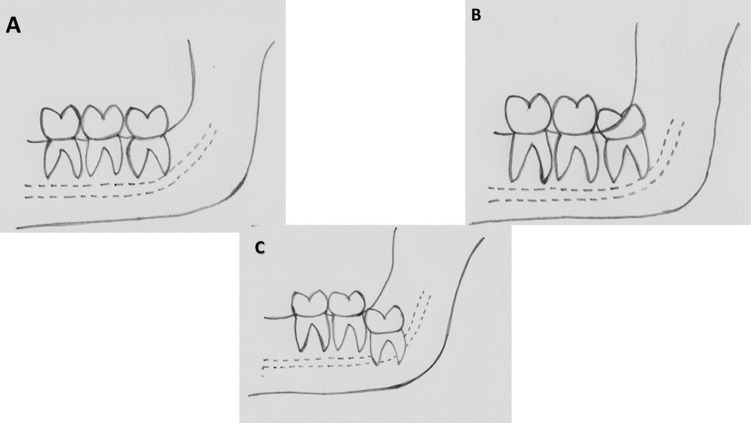
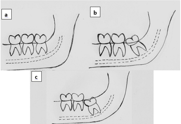
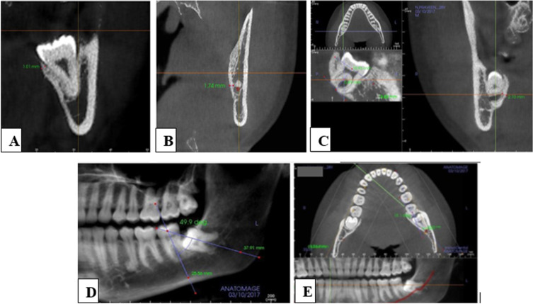
Methods: This was a retrospective study on CBCT images of 648 mandibular third molars. The lingual plate thickness at three different root levels - cervical, mid-root, and apex along with the position of the tooth, number of roots, density of lingual plate, age, and gender were evaluated. The measurements were done on Invivo 5-Anatomage software. Statistical comparison of the categorical variables was done by Chi-square test, and Fisher's exact test, and univariate and multivariate analysis were done using binomial logistic regression.
Results: Lingual plate thickness of the third molars at the cervical, mid root, and apex were 1.28 mm, 1.42 mm and .01 mm (mean). A significantly higher proportion of subjects with thin lingual plates at mid-root (p-value=0.01) and apex (p-value=0.05) were in the 21-30 age group. Lingual bone density was significantly associated with the thickness of the lingual plate at the mid-root. A significantly higher proportion of thinner lingual plates at the mid-root level were associated with mesioangularly placed third molars (p-value=0.002).
Conclusion: Our study presented that lingual plate thickness has a strong association with age, angulation, and the number of roots. Knowledge about these risk factors is imperative during the management of third molar impactions.


 分享
分享
 求助内容:
求助内容: 应助结果提醒方式:
 扫码关注我们
